Whole slide imaging, the technology that turns glass microscope slides into high-resolution digital images that can be viewed from anywhere, has taken another small but meaningful step toward routine use in thyroid diagnostics. In a retrospective pilot study conducted at two Turkish hospitals, pathologists examining digitized thyroid cytology slides remotely agreed with the conventional glass-slide diagnosis in 69.2 percent of cases, with statistical measures of agreement falling in the moderate to substantial range. The study, published in Diagnostic Pathology, offers preliminary evidence that remote digital diagnosis of thyroid fine-needle aspiration specimens is technically feasible even in settings without dedicated digital pathology infrastructure, while also highlighting exactly where the technology struggles.
The research team, led by pathologists at Malatya Turgut Özal University Training and Research Hospital together with colleagues at the University of Health Sciences Turkey, Istanbul Prof. Dr. Cemil Taşcıoğlu City Hospital, assembled thirteen thyroid cases retrospectively from routine fine-needle aspiration specimens evaluated between June 2025 and February 2026. The cohort was deliberately constructed to span the diagnostic spectrum encountered in clinical practice, including benign lesions, malignant tumors, indeterminate categories, and one low-risk tumor. The patients reflected the typical demographic of thyroid nodular disease: twelve of the thirteen were women, and the mean age was 47.5 years, with a range of 30 to 60 years. Most nodules showed heterogeneous or multinodular morphology on ultrasound, frequently accompanied by cystic components, and hypoechogenicity was the most common ultrasonographic feature.
The study design was straightforward but rigorous in its blinding. One board-certified pathologist established the reference diagnosis by examining the glass slides with a conventional light microscope on-site. Two independent pathologists then evaluated the same cases using whole slide images, working remotely and without access to the reference diagnosis or to each other’s interpretations. To mirror real clinical conditions, the digital reviewers were given the biopsy identification number, patient age, sex, radiological findings, and thyroid function test results, the same information a working pathologist would normally have. The conventional glass-slide diagnosis served as the primary reference standard, a choice the authors themselves flag as a potential source of bias, since it reflects routine clinical diagnosis rather than an independent gold standard.
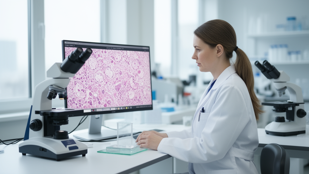
The logistics of the study reveal much about the practical barriers to digital pathology in resource-limited settings. The primary institution had no in-house whole slide scanner, so glass slides had to be physically transported by medical courier roughly 1,100 kilometers from Malatya to Istanbul for digitization, a journey taking about a day, before being returned under controlled conditions. In total, 45 slides stained with hematoxylin and eosin, Papanicolaou, or Giemsa were transferred and evaluated, with approximately four to six representative slides per case. All slides were scanned with a Hamamatsu NanoZoomer S360 whole-slide scanner at 20-times magnification, without Z-stacking, the multi-focal plane acquisition that cytology specimens often require because of their three-dimensional architecture. The resulting digital files, typically between 2 and 3 gigabytes per slide, were too large for direct transfer, so the pathologists accessed them remotely through the AnyDesk desktop-sharing platform, with each session individually authorized and supervised by technical staff to protect patient confidentiality.
The results painted a nuanced picture. Each of the two remote observers matched the reference diagnosis in 9 of 13 cases, yielding the overall concordance rate of 69.2 percent. Cohen’s kappa, a statistic that measures agreement beyond what would be expected by chance, was approximately 0.63 for one pathologist and 0.58 for the other, values conventionally interpreted as moderate to substantial agreement. However, the confidence intervals were wide, spanning from 0.33 to 0.93 for one observer and 0.28 to 0.88 for the other, a statistical uncertainty the authors attribute directly to the small sample size and emphasize repeatedly as a reason for caution.
Perhaps the most reassuring finding concerned the clinically decisive categories. Full concordance among all observers was observed in 7 cases, predominantly malignant diagnoses, which were mostly papillary thyroid carcinoma. Partial concordance occurred in 5 cases, mainly involving borderline or intermediate diagnostic groups where interpretations shifted slightly between adjacent categories. Complete discordance appeared in just one case, within an indeterminate cytological category. Crucially, no direct discordance between clearly benign and malignant diagnoses was observed anywhere in the cohort, and the authors report no clinically significant misclassification between benign and malignant categories. Discrepancies, when they occurred, took the form of shifts between neighboring diagnostic groups, such as benign to nondiagnostic, atypia of undetermined significance to follicular neoplasm, or suspicious to malignant.
This pattern of error is significant because it mirrors a well-known feature of thyroid cytopathology itself rather than a flaw specific to digital imaging. The Bethesda System for Reporting Thyroid Cytopathology has greatly standardized thyroid reporting, but categories such as atypia of undetermined significance and suspicious for malignancy remain diagnostic grey zones with considerable interobserver variability and heterogeneous malignancy risk, even when pathologists use conventional microscopes. The study’s final diagnoses comprised six malignant lesions, three benign lesions, one low-risk tumor classified as suspicious for a follicular neoplasm, and three indeterminate cytological categories, and it was precisely within these indeterminate groups that most discrepancies clustered. The authors argue that the variability observed should be interpreted within the broader context of cytopathology rather than attributed solely to the digital modality.
The technical challenges of digitizing cytology, as opposed to histopathology, also loom large in the findings. Cytological preparations exhibit complex three-dimensional architecture, with overlapping cellular clusters and variable thickness, and optimal visualization often requires multi-focal Z-stacking acquisition, which increases scanning time, data storage demands, and computational burden. Because this study used single-plane scanning, the authors acknowledge that the absence of Z-stacking may have contributed to diagnostic uncertainty in certain cases. The choice of stains, including H&E, Papanicolaou, and Giemsa preparations evaluated according to case availability, reflected real-world practice but added another layer of variability. These constraints, combined with the delays and file-size hurdles of the courier-based workflow, may have influenced interpretative conditions, although the authors describe the desktop-sharing approach as a pragmatic, cost-effective, and reliable alternative where dedicated infrastructure is unavailable, while stressing that it should be considered a transitional solution rather than a sustainable routine model.
The wider context makes the study’s modest size easier to understand. Whole slide imaging enables remote access to cases, telepathology consultation, digital archiving, and support for multidisciplinary discussions, and it serves as critical infrastructure for future artificial intelligence-driven decision support in cytopathology. For peripheral or resource-limited centers, the ability to have cases reviewed by subspecialty experts across institutions could be especially valuable. Yet adoption in middle-income countries is often constrained by infrastructural, economic, and regulatory limitations and by the lack of locally generated validation data, and the College of American Pathologists continues to emphasize the need for rigorous validation studies with adequate case numbers before routine clinical implementation. By conducting a structured validation aligned with these recommendations in a real-world setting, the Turkish team addressed a documented gap in the literature on digital pathology in emerging healthcare systems.
The authors are careful about what their data can and cannot show. Beyond the small sample and the use of conventional cytology rather than histopathology as the uniform reference standard, they note that histopathological outcomes were available only for a subset of cases and were used solely for secondary descriptive analysis. The wide confidence intervals around the kappa estimates indicate substantial statistical uncertainty, and the findings are explicitly framed as preliminary. Their conclusion is measured: whole slide imaging demonstrated moderate agreement with conventional microscopy in well-defined thyroid cytopathology categories, variability was more pronounced in indeterminate cases reflecting inherent diagnostic challenges, and the results support the preliminary feasibility of remote digital workflows. Confirming that promise, they write, will require larger, multicenter validation studies with standardized protocols before digital thyroid cytology can move from pilot studies into everyday practice.
Subject of Research: Diagnostic concordance between remote whole slide imaging and conventional microscopy in thyroid cytopathology
Article Title: Diagnostic concordance of remote WSI-based thyroid cytology compared to conventional cytological diagnosis: a pilot study
Article References: Şahi̇n, A. F., Yalçin, Ö., Büyücek, S., Dogan Ayan, M., & Elmas, H. (2026). Diagnostic concordance of remote WSI-based thyroid cytology compared to conventional cytological diagnosis: a pilot study. Diagnostic Pathology, 21(1), Article 66. https://doi.org/10.1186/s13000-026-01820-9
Image Credits: AI Generated
DOI: 10.1186/s13000-026-01820-9
Keywords: whole slide imaging, digital pathology, thyroid cytology, fine-needle aspiration, telepathology, Bethesda System, diagnostic concordance, Cohen's kappa, pilot study, remote diagnosis, cytopathology, papillary thyroid carcinoma
News Source: Ophelia Keating. (October 29, 2026). Remote Digital Slides Match Microscope Diagnosis in Most Thyroid Cytology Cases. Scienmag.