


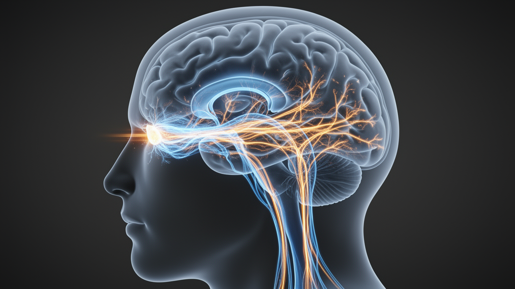
Acquired cortical blindness has long been treated as a one-way door. When the primary visual cortex — the brain region that decodes signals arriving from the eyes — is damaged by stroke or trauma, patients are traditionally told that conscious vision is gone for good. A new systematic review published in the Egyptian Journal of Neurosurgery challenges that fatalism, but with a caveat that is as important as the headline finding: the evidence that vision can be partially retrained comes from just three small, non-randomized studies, and the authors themselves rate the certainty of that evidence as low.
The review, led by Karen Dayana Villarreal Marín and colleagues, followed PRISMA 2020 guidelines and searched seven databases, including PubMed/MEDLINE, Web of Science, Scopus, ScienceDirect, SciELO, ClinicalKey and Google Scholar, for studies published between 2021 and 2025 in English or Spanish. The search retrieved a staggering 15,245 records. After removing duplicates, screening titles and abstracts, and assessing full texts against strict eligibility criteria, only three studies survived. The authors attribute this dramatic attrition to the combination of a deliberately broad initial search and narrow criteria requiring explicit, quantifiable evaluation of both brain plasticity mechanisms and residual visual function in acquired, non-congenital cortical blindness.
The three included studies — two quasi-experimental intervention studies and one longitudinal neuroimaging study — were conducted in the United States and Canada and enrolled adults with cortical blindness caused by occipital lobe lesions of ischemic or hemorrhagic origin. In all three, targeted training was associated with activation of perilesional residual activity in the primary visual cortex, subcortical collicular pathways, and extrastriate circuits, producing improvements in luminance detection, visual field recovery, and generalization of training gains to untrained stimuli.
The largest cohort came from Barbot and colleagues, who studied eleven adults (mean age 61 years) with chronic, unilateral, stroke-induced cortical blindness. All had damage in the posterior cerebral artery territory, with lesions concentrated in the medial occipital lobe and calcarine sulcus, and lesion volumes ranging enormously from 667 to 64,320 cubic millimetres. Crucially, these patients were far beyond the typical six-month window of spontaneous recovery — their injuries dated from 5 to 237 months earlier. The study reported that the amount of spared perilesional V1 activity, measured with retinotopic functional MRI, was associated with the magnitude of training-induced recovery in luminance sensitivity, with enlargement of population receptive fields in perilesional cortex proposed as the imaging correlate. The review’s authors stress, however, that this remains an imaging-behaviour association rather than proof of a causal mechanism.
Rowland and colleagues took a different route, enrolling just two men with chronic stroke-induced homonymous hemianopia — a partial field loss that the review carefully distinguishes from complete cortical blindness. After multisensory visual-auditory training, overt detection was reportedly restored across the entire hemianopic field within weeks. The proposed explanation involves re-sensitization of superior colliculus output neurons via a pathway running through the ventrolateral pulvinar to extrastriate cortex. But the review is emphatic that this mechanistic account is a plausible interpretation drawn from the wider multisensory-integration literature: Rowland’s team never directly measured subcortical activity in their patients. With two participants, no control condition, and no possibility of blinding, the study received a critical risk-of-bias rating.
The third study, by Awada and colleagues in Canada, screened 37 patients and trained seven with complex optic flow stimuli — the expanding visual patterns generated when moving through space. Four trained regularly, at five or more sessions per week, while three trained sporadically, a difference in effective intervention dose that the review flags as a significant confound. The study reported broader spatial generalization of recovery with complex stimuli than simpler ones, a pattern the authors propose is consistent with engagement of a direct pathway from the lateral geniculate nucleus to the motion-specialized hMT+ cortex and the MST area. Again, the review notes this pathway-level account is inferred from the broader neuroscience literature, not directly measured in Awada’s dataset. Notably, lesion characteristics were not systematically described for individual participants, a reporting limitation that precluded any analysis of how structural damage modulated training response.
Methodological quality was the review’s sobering centerpiece. Using the ROBINS-I tool, all three studies showed confounding as their most vulnerable domain, reflecting the ethical and logistical difficulty of running randomized controlled trials in this population. Barbot et al. fared best with moderate risk, aided by blinded evaluators and objective retinotopic fMRI outcomes; Awada et al. received serious risk due to the absent control group and variable adherence; Rowland et al. was rated critical. On the SIGN classification, all three studies sat at evidence level 2+ with recommendation grade C, and GRADE assessment judged the overall certainty of evidence for plasticity-based rehabilitation to be low — meaning the true effect may differ substantially from what is reported.
What the review does defend is a conceptual shift. The authors argue the findings are consistent with a dynamic model in which recovery potential reflects an interaction between lesion anatomy and the integrity of subcortical and extrastriate visual circuits, rather than being determined by V1 damage alone. This aligns with the broader phenomenon of blindsight — the ability of some patients to respond to visual stimuli without conscious perception — and with evidence that perilesional and extrastriate networks can remain functionally active or be modulated through specific interventions. Even so, the authors concede that a three-study qualitative synthesis cannot establish this model definitively, and that no meta-analysis was possible given the clinical and methodological heterogeneity of the included work.
The review also identifies conspicuous gaps. None of the three studies used diffusion tensor imaging tractography to characterize white-matter integrity, none stratified outcomes by lesion profile, and none incorporated neuromodulation. Evidence from outside the included studies suggests that anodal transcranial direct current stimulation over occipital cortex, combined with perceptual training, may accelerate detection recovery in hemianopia patients, and that intact LGN–hMT+ pathways are associated with better transfer of training gains — but the review explicitly presents these as testable hypotheses for future trials, not as extensions of its own results. There is also a geographic problem: nearly all research comes from high-income countries, leaving patients in Latin America and other resource-limited settings, where the burden of stroke-induced cortical blindness is substantial, largely outside the evidence base.
For clinicians, the practical message is deliberately restrained. The authors suggest that measurable residual visual function, or its potential to be unmasked, argues for early referral to visual neurorehabilitation once patients are medically stable, and that functional imaging could in principle help identify likely responders — though that application has not been tested prospectively. Above all, they urge individualized, modest expectations: lesion volume, laterality, chronicity and subregion involvement varied more within each study than the interventions varied across them, and current evidence does not support any single rehabilitation protocol as a standard of care. The review was not prospectively registered, an acknowledged limitation, and no inter-rater agreement statistic was calculated. The bottom line is genuinely two-sided: the chronically injured visual cortex may retain more trainable capacity than classic neuroanatomy assumed, but with three small, bias-prone studies underpinning that claim, the field’s most important finding may be how much it still needs to find out.
Subject of Research: Brain plasticity and residual visual function in adults with acquired cortical blindness
Article Title: Brain plasticity and residual visual functions in acquired cortical blindness: a systematic review of the literature
Article References: Villarreal Marín, K. D., Gil, S. L. L., Gil, L. V. L., Bonilla, A. K. O., Cuellar, J. D. C., & Novoa Álvarez, R. A. (2026). Brain plasticity and residual visual functions in acquired cortical blindness: a systematic review of the literature. Egyptian Journal of Neurosurgery, 41(1), Article 169. https://doi.org/10.1186/s41984-026-00677-w
Image Credits: AI Generated
DOI: 10.1186/s41984-026-00677-w
Keywords: cortical blindness, brain plasticity, blindsight, visual rehabilitation, primary visual cortex, systematic review, neuroimaging, stroke, hemianopia, neurorehabilitation, risk of bias, optic flow
News Source: Cassandra Pierce. (October 29, 2026). Blind but Not Blind to Hope: Training May Reroute Vision After Cortical Blindness. Scienmag.



