


Pneumonia is the commonest single infection leading to intensive care admission and the commonest nosocomial infection acquired while patients are in the intensive care unit. A new commentary in Intensive Care Medicine, written by Andrew Conway Morris, Shyamanova Mazumdar and Mark Jeffrey, now argues that the hidden differences are not peripheral details but central to how the condition should be understood and, eventually, treated.
For years, the underlying histological picture of pneumonia has typically been assumed to be neutrophil-dominant consolidation, with or without diffuse alveolar damage and hyaline membranes, the hallmark of acute respiratory distress syndrome. At the same time, the identification of reproducible peripheral blood phenotypes in ARDS and in respiratory sepsis has fuelled renewed efforts towards stratified medicine in these syndromes. The commentary raises a fundamental problem with that approach: if inflammation in the blood does not reflect what is happening in the lung, therapies selected on the basis of blood tests may simply be applied to the wrong patients.
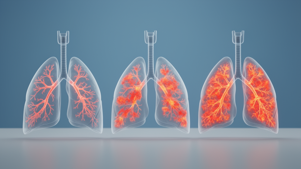
The authors draw on their own recent work sub-phenotyping the lungs of four cohorts of patients with severe pneumonia. Using transcriptomic analysis of cells obtained by bronchoalveolar lavage, or BAL, from patients with suspected pneumonia, the team identified three distinct pulmonary phenotypes, which they termed pneumotypes: Pneumotype 1, 2 and 3. These can be summarised as an immunologically silent pattern, a balanced immune response, and a hyperinflammatory pattern dominated by immature neutrophils. Each pneumotype carried a distinct cellular and molecular signature, and the commentary sets out the inferred pathophysiological mechanisms behind each, including altered surfactant processing, epithelial repair programmes and self-reinforcing inflammatory cycles.
The compartmentalisation of these phenotypes is the most consequential finding. The pneumotypes were not detectable in the blood, and re-analysis suggested this absence was not a matter of sample size but a genuine lack of biological signal in the circulation. Blood transcriptomic analysis did reveal two sub-phenotypes, closely related to the previously described Sepsis Response Signatures, but these blood patterns did not map onto the lung patterns. In other words, the lung and the blood were telling two different stories about the same patient, and neither could be read as a proxy for the other.
The clinical implications of that mismatch are stark. When the researchers examined interactions between pneumotype and blood type in predicting mortality, the worst outcomes were found in patients who had the least inflammation in their lungs but carried the more inflammatory blood signature. This is precisely the sort of discordance that earlier work had flagged as a source of concern for blood-based stratification. A patient whose circulation screams inflammation may, at the alveolar level, have an almost silent process, and treating the blood picture rather than the lung picture could misdirect therapy entirely.
The first pneumotype, Pn1, is the most conceptually disruptive. It presents a hypoinflammatory picture with low alveolar cytokine levels, scanty neutrophils and expanded alveolar macrophages. Although the phenotype was enriched for patients with immunosuppression, 44 percent of patients with this pneumotype were not immunosuppressed, so immunosuppression alone does not explain it. Despite its quiet inflammatory profile, Pn1 was associated with a similar severity of respiratory failure to the other phenotypes and with high rates of alveolar haemorrhage. Together with elevated epithelial damage markers, this points to what the authors describe as an immunologically silent disruption of the alveolar epithelial barrier, severe lung injury occurring with almost no classical inflammatory response.
The molecular clues in Pn1 converge on surfactant metabolism. Increased expression of phospholipase A2, encoded by PLA2G15, implies increased breakdown of the surfactant phosphatidylcholine, while reduced expression of lysophosphatidylcholine acyltransferase 1, or LPCAT1, points to a failure to recycle the hydrolysed lipid. The predicted consequence is an accumulation of tissue-toxic lysophosphatidylcholine that damages the delicate alveolar epithelium. At the same time, increased expression of CD36, a scavenger receptor macrophages use to remove lipids including surfactant, suggests the formation of lipid-laden alveolar macrophages, a feature seen in several other pulmonary inflammatory diseases. Tissue Factor, encoded by F3, was also reduced in expression; in a mouse lipopolysaccharide model, F3 deletion increases pulmonary haemorrhage and lipid oxidation with a macrophage-dominant infiltrate and modest cytokine levels, although the authors stress that these mechanisms require further exploration. The prominence of abnormal surfactant processing in the most common pneumotype may, they suggest, explain why previous trials of exogenous surfactant in ARDS failed despite widely described surfactant deficits in patients.
At the opposite end of the spectrum, Pn2, the balanced phenotype, carried the best prognosis. It occupies an immunologically intermediate position, with moderate concentrations of alveolar cytokines and moderate numbers of neutrophils. What distinguished it most strikingly was a prominent epithelial repair signature and increased expression of stem cell genes, notably SOX2 and SOX9. In the single-cell dataset used for validation, the team identified a population of distal alveolar stem cells in Pn2 patients, which may be driving the repair phenotype. Consistent with a regenerative program, Pn2 patients had the fastest time to ventilator liberation and the lowest mortality of the three groups. In animal models of respiratory infection, application of these distal alveolar stem cells has been reported to improve survival, although translating that observation into human therapy remains distant.
Pn3, the hyperinflammatory phenotype, offers a mechanistic account of a familiar clinical frustration: inflammation that persists after the pathogen is gone. Patients given appropriate antibiotics for pneumonia usually clear the infection rapidly, yet lung inflammation and injury often continue, driving ongoing or escalating antimicrobial therapy. In Pn3, elevated cytokines coincide with prominent neutrophil and monocyte infiltration, and the researchers found genetic signatures of neutrophil immaturity, including increased expression of IL1R2, ARG1 and PADI4, which validation in a single-cell dataset confirmed. Immature neutrophils are both immunoparetic and have impaired antimicrobial function. Although the lung phenotypes are compartmentalised, pulmonary and blood concentrations of interleukin-6 and granulocyte-stimulating factor did correlate, and both are potent stimuli for emergency granulopoiesis, the accelerated production and release of immature neutrophils by the bone marrow. The lung recruits immature neutrophils, the marrow responds to the cytokine signal, and the two loops reinforce each other, potentially becoming locked on and sustaining injury even after the initial precipitant has been removed. Notably, Pn3 was not synonymous with the maladaptive Sepsis Response Signature 1, suggesting selective recruitment of immature neutrophils to the lungs rather than a simple mirror of the blood.
The authors are careful about what stands between these findings and the bedside. Translating the pneumotypes into routine clinical use will require a clinically usable classifier, for example a parsimonious gene expression signature enabling rapid near-bedside identification of pneumotypes alongside pathogen detection. The current work reflects a snapshot in time, so serial sampling studies will be needed to establish how stable the pneumotypes are over the course of illness and where the therapeutic windows lie. The derivation work used formal, high-volume BAL, but validation indicated that mini-BAL may be able to discriminate the phenotypes, and the possibility of a less invasive test, if the signatures could be detected outside the lung, remains open. For Pn3, repurposing existing licensed therapeutics such as interleukin-6 receptor antagonists to interrupt the self-reinforcing signalling cycle is described as the fastest route, though it would require stratified trials using the proposed classifier; for the other pneumotypes, further confirmation of the targets is needed first. What the commentary establishes now is a conceptual shift: pneumonia in the critically ill is not one disease with one histology, and stratified therapies that focus purely on the blood compartment may miss the most important nuances of what is happening in the lung itself.
Subject of Research: Compartmentalised lung inflammation sub-phenotypes in severe pneumonia and their implications for critical care
Article Title: What do the findings of compartmentalised sub-phenotypes in lungs of patients with pneumonia mean for critical care?
Article References: Conway Morris, A., Mazumdar, S., & Jeffrey, M. (2026). What do the findings of compartmentalised sub-phenotypes in lungs of patients with pneumonia mean for critical care?. Intensive Care Medicine. https://doi.org/10.1007/s00134-026-08637-6
Image Credits: AI Generated
DOI: 10.1007/s00134-026-08637-6
Keywords: pneumonia, ARDS, pneumotypes, bronchoalveolar lavage, transcriptomics, neutrophils, surfactant, alveolar epithelium, IL-6, sepsis response signatures, stratified medicine, critical care
News Source: Ophelia Keating. (October 29, 2026). Three lung inflammation sub-phenotypes in pneumonia may reshape critical care treatment. Scienmag.



