


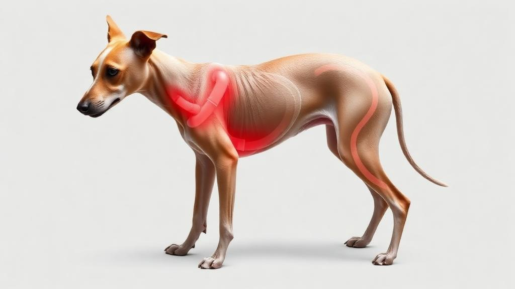
Whippets, the sleek sprinting sighthounds beloved of racing enthusiasts, carry a hidden anatomical signature that shows up clearly on computed tomography scans. A retrospective study of 68 dogs has found that whippets have significantly larger abdominal caudal vena cava to aorta ratios (CVC/Ao) than other dogs, while their thoracic ratios are indistinguishable from those of non-whippet controls. The finding, published in Frontiers in Veterinary Science, suggests that a measurement veterinarians routinely use to gauge fluid status may need breed-specific interpretation, at least in this athletic breed.
The caudal vena cava to aorta ratio is an established index of intravascular volume status. The ratio was first described in human paediatrics on ultrasound to assess fluid status in emergency settings and was later extrapolated to canine patients. It can be assessed both ultrasonographically and on CT, at thoracic and abdominal sites, but breed-specific values had never been reported before this study.
Researchers at Dick White Referrals in Cambridgeshire, United Kingdom, searched their clinical database for whippets that had undergone CT between October 2011 and July 2025 for reasons unrelated to suspected cardiovascular disease. They identified 34 whippets and randomly selected 34 non-whippet control dogs scanned at the same body regions and without known cardiovascular disease. All CT studies were performed under general anaesthesia in sternal recumbency, with measurements taken at standardised thoracic and abdominal sites by an observer who was not blinded to breed. A second, blinded observer re-measured a random subset of 20 dogs to assess reliability.
The results were striking. Whippets had a mean abdominal CVC/Ao of 1.54 plus or minus 0.29, compared with 1.28 plus or minus 0.23 in controls, a mean difference of 0.26 (95% confidence interval 0.13 to 0.39; p < 0.001; Cohen’s d = 0.99). At the thoracic site, however, the two groups were statistically indistinguishable: 1.22 plus or minus 0.23 in whippets versus 1.28 plus or minus 0.26 in controls (p = 0.29). Because whippets were both lighter (mean 14.3 kg versus 22.2 kg) and younger (mean 6 years 4 months versus 8 years 4 months) than the controls, the team also ran an analysis of covariance adjusting for weight and age. The abdominal difference not only persisted but grew slightly, with an adjusted difference of 0.29 (95% CI 0.15 to 0.42; p < 0.001), and restricting the analysis to the weight range common to both groups gave near-identical results.
A within-animal comparison reinforced that the elevation is regional rather than generalised. In whippets, the abdominal ratio exceeded the thoracic ratio by a mean of 0.32 (95% CI 0.19 to 0.46; p < 0.001), whereas in controls the two segments were essentially identical (mean difference 0.00; p = 0.995). The authors speculate that anatomical constraints within the thorax, where the diaphragm, mediastinum and right atrium may limit how far the cava can dilate, could explain why increased venous return is accommodated more readily in the abdomen, though they stress this idea is extrapolated from human data and remains unconfirmed in dogs.
The study also delivered a cautionary message about published reference values. Both groups exceeded the most recent CT-derived values from anaesthetised dogs of various breeds and clinical conditions (thorax 1.12 plus or minus 0.23; abdomen 0.78 plus or minus 0.25) at both sites. Compared with the ultrasonographic reference interval for healthy conscious dogs (0.93 to 1.32), 25 of 34 whippets (73.5%) and 14 of 34 controls (41.2%) lay above the upper limit of 1.32, a significantly greater proportion in whippets (p = 0.007). The authors conclude that directly applying ultrasonographic reference values from conscious dogs to anaesthetised CT patients, or CT-derived ratios that disregard breed, is not recommended.
To give clinicians something practical to work with, the team derived preliminary mean plus or minus two standard deviations limits: 0.96 to 2.13 for the abdominal ratio in whippets, 0.82 to 1.75 for abdominal ratios in controls, and a single combined thoracic limit of 0.76 to 1.74 across all 68 dogs. They are careful to frame these as cohort observations rather than formal breed-specific reference ranges, since the sample of 34 per group falls below the ASVCP-recommended minimum of 120 and the limits were derived parametrically rather than by the preferred non-parametric method.
The authors also flag two interpretive caveats. An elevated ratio could arise from a larger caudal vena cava, a smaller aorta, or both, and the study reports only the ratio, so whether the difference reflects greater venous capacitance or breed-related aortic dimensions remains undetermined. And although one control dog happened to be a greyhound, another sighthound, its abdominal ratio of 1.33 sat close to the control group mean, raising the possibility that the finding is specific to whippets rather than common to sighthounds generally, though a single animal permits no inference.
Several limitations temper the conclusions. The retrospective design meant anaesthesia protocols were not standardised, two different CT scanners were used unevenly across the groups, and the breath-hold technique differed largely along group lines, although the authors argue the direction of these biases would tend to narrow rather than widen the observed difference. Dogs were not excluded for intra-abdominal disease, which can alter caval diameter, but pressure-relevant abdominal lesions were if anything more frequent in controls. The initial measurements were taken by an observer aware of group assignment; blinded re-measurement showed excellent interobserver agreement for the thoracic ratio (ICC 0.96) but only moderate agreement for the abdominal ratio (ICC 0.67), with a systematic difference of about 0.13 that was, importantly, of similar magnitude in both groups. The ratio also does not fully normalise for body size, since body weight remained positively associated with CVC/Ao at both sites, and the thoracic ratio declined with age independently of breed and weight (minus 0.023 per year; p = 0.002).
Clinically, the practical takeaway is narrow but useful: an abdominal CVC/Ao above the published ultrasonographic upper limit should not, on its own, be interpreted as evidence of volume overload or right-sided heart disease in an anaesthetised whippet. The mechanism underlying the regional difference remains to be established, and the authors call for prospective studies in targeted breed groups using standardised CT acquisition and anaesthetic protocols, including blood pressure and echocardiographic data, along with studies reporting absolute vessel dimensions indexed to body size. Until then, the whippet’s unusually roomy abdominal vein stands as a reminder that even well-worn veterinary measurements can carry breed-specific surprises.
Subject of Research: Breed differences in CT-measured caudal vena cava to aorta ratios in whippets versus other dogs
Article Title: Whippets have larger abdominal caudal vena cava to aorta ratios than other dogs on computed tomography
Article References: Esmieu, S. L., Ma, M.-H., Mantis, P., Biondo, S., Constantinescu, R., & Furtado, A. R. R. (2026). Whippets have larger abdominal caudal vena cava to aorta ratios than other dogs on computed tomography. Frontiers in Veterinary Science, 13, Article 1971485. https://doi.org/10.3389/fvets.2026.1971485
Image Credits: AI Generated
DOI: 10.3389/fvets.2026.1971485
Keywords: whippets, caudal vena cava, aorta, computed tomography, veterinary radiology, reference intervals, sighthounds, intravascular volume, dogs, general anaesthesia, breed differences, veterinary imaging
News Source: William Thompson. (October 29, 2026). Research report: Whippets have larger abdominal caudal vena cava to aorta ratios than other dogs on computed tomography. Scienmag.



