


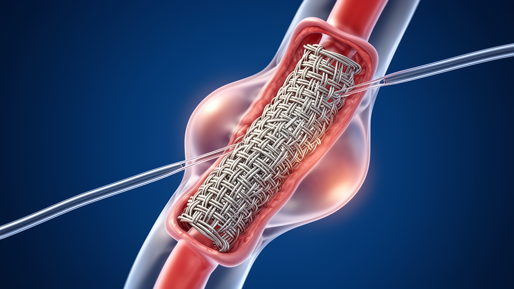
The procedure, known as physician-modified fenestrated/inner-branched endovascular aortic repair, or PM-F/iBEVAR, sealed the leak that had driven the aneurysm’s enlargement and preserved blood flow to all three major branches of the aortic arch. Six months later, the aneurysm had not grown further, although important questions about long-term durability remain open.
The story began four years before the repair, when the patient developed an uncomplicated DeBakey type IIIa acute aortic dissection, a tear in the inner layer of the body’s main artery with the entry point located in the distal aortic arch. He was initially managed with conservative follow-up, but a year later he suffered a cerebral infarction that was highly suspected to be an embolic event related to the dissection. Subsequent computed tomography revealed persistent blood flow within the false lumen, the false channel created when the arterial wall splits, and the distal aortic arch had dilated to more than 40 millimeters.
The proximal landing zone diameter measured approximately 27 millimeters, so a 30-millimeter graft was chosen, giving an oversizing rate of roughly 11 percent. During follow-up, however, imaging showed trouble. A CT scan performed in the referral year revealed persistent blood flow within the proximal false lumen caused by a type Ia endoleak, meaning blood was leaking around the top of the graft and back into the aneurysm sac. The distal aortic arch diameter had grown from 33 millimeters two years earlier to 44 millimeters, an aneurysmal degeneration that prompted referral for further intervention.
The patient’s medical history made standard surgery a poor option. He had accumulated multiple cerebral infarctions, an intracerebral hemorrhage, and renal dysfunction, and he lived with residual right-sided limb ataxia and dysarthria. These neurological sequelae impaired his activities of daily living, and after multidisciplinary discussion the aortic team judged conventional arch replacement unsuitable. The team weighed alternatives carefully. Zone 2 TEVAR with either one-vessel debranching or left subclavian artery fenestration was considered, but centerline analysis showed the proximal sealing zone measured only 18 millimeters, which the authors deemed inadequate for a secure seal. A commercially designed thoracic branch endoprosthesis intended for zone 2 landing was rejected for the same anatomical reason, and debranching would have required an additional bypass operation with a risk of graft occlusion.
Other endovascular workarounds also fell short in this anatomy. A zone 1 landing was judged too difficult because the brachiocephalic artery and left common carotid artery originated from almost the same location, leaving an extremely short inter-vascular distance. Retrograde in situ branched stent-grafting and in situ fenestration, techniques that create branch access during the operation itself, offer the advantage of requiring no pre-procedural modification and being immediately available in emergencies, but they carry concerns about cerebral embolism, type IIIc endoleaks, and damage to the vessel wall or graft material during fenestration. In this patient, the acute angulation at the origin of the left subclavian artery made both approaches technically unattractive. Commercially available multibranched or patient-specific custom-made grafts for zone 0 landing are not yet approved in Japan, leaving physician modification as the remaining route to a zone 0 repair.
The entire modification took 55 minutes.
Final angiography showed no endoleak. The operation lasted 115 minutes, with an estimated blood loss of 100 milliliters, radiation exposure of 512 milligray, and contrast volume of 95 milliliters.
The postoperative course was largely reassuring. The patient was discharged without complications. At the six-month follow-up, contrast-enhanced CT was omitted, so the presence of an endoleak could not be evaluated; however, plain CT showed the supra-aortic branches still patent and the aneurysm diameter at 42 millimeters, with no significant interval change.
The authors place the case in a broader context of risk. The predicted operative mortality by the JapanSCORE was 5.3 percent, and the patient’s modified Rankin Scale score was grade 3 or 4, reflecting moderate to severe disability; the decision for endovascular repair rested on his cumulative vulnerabilities rather than the score alone. They attribute the original migration to the non-bare-stent design and the steep arch, factors previously associated with bird-beak formation and migration.
The report comes with explicit limitations. Long-term durability and safety of PM-F/iBEVAR remain unclear, endoleaks may still appear and require secondary intervention, and zone 0 repair may raise the risk of retrograde type A dissection. Because contrast-enhanced CT was skipped at six months, a persistent or recurrent endoleak could not be definitively excluded, and the type II leak, though possibly self-limiting, still requires surveillance. The authors also emphasize that PM-F/iBEVAR is a technically demanding, off-label procedure reserved for carefully selected patients at experienced aortic centers with appropriate institutional and ethical approval. Previous reports of the technique for distal arch aneurysms have shown favorable short-term outcomes, including high branch patency and freedom from reintervention, and this case adds evidence that it can serve as a rescue strategy after TEVAR failure caused by stent graft migration.
Subject of Research: Physician-modified fenestrated/inner-branched endovascular aortic repair for distal aortic arch aneurysm
Article Title: Physician-modified fenestrated/inner-branched endovascular aortic repair for distal aortic arch aneurysm after stent graft migration: a case report
Article References: Hori, Y., Shibata, T., Tsushima, S., Yoshikawa, K., Hayasaka, S., Sugiura, H., Maeda, H., Nakajima, T., Nakazawa, J., Arihara, A., Kato, K., Komatsu, S., Yonemori, M., & Iba, Y. (2026). Physician-modified fenestrated/inner-branched endovascular aortic repair for distal aortic arch aneurysm after stent graft migration: a case report. General Thoracic and Cardiovascular Surgery Cases, 5(1), Article 40. https://doi.org/10.1186/s44215-026-00272-3
Image Credits: AI Generated
DOI: 10.1186/s44215-026-00272-3
Keywords: aortic arch aneurysm, endovascular repair, PM-F/iBEVAR, TEVAR, stent graft migration, endoleak, case report, physician-modified stent graft, zone 0 landing, aortic dissection, Physician-modified, fenestrated
News Source: Ophelia Keating. (October 29, 2026). Custom-Modified Stent Graft Rescues Aneurysm After Failed Aortic Repair. Scienmag.



