


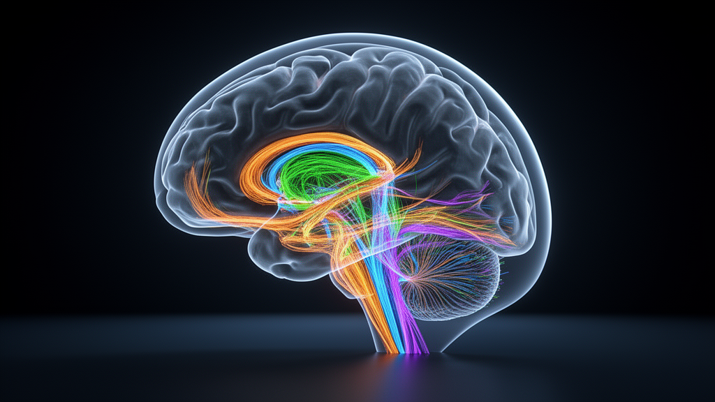
Deep inside each cerebral hemisphere, a narrow band of white matter funnels traffic between the temporal lobe, the frontal lobe and the diencephalon. Neurosurgeons call it the temporal stem, and for decades its exact wiring has been contested territory. Now a team of researchers has produced one of the most complete three-dimensional maps of this structure to date, using diffusion tensor tractography to digitally dissect twenty human brains from the publicly available Human Connectome Project dataset. Their analysis, published in the journal Discover Imaging, identifies six distinct fibre tracts within the temporal stem and establishes, for the first time in a single tractographic study, their relative sizes, layered arrangement and spatial relationships.
The temporal stem is bounded inferiorly by the roof of the temporal horn of the lateral ventricle and superiorly by the inferior limiting sulcus, with the amygdala marking its anterior limit and the lateral geniculate nucleus its posterior boundary. Despite more than twenty years of intensive research, no consensus has emerged on the morphological and topographic anatomy of the fibre tracts that pass through it, or on a clear depiction of their spatial relationships. The authors of the new study note that a comprehensive literature review revealed no previous work describing the dissection of all six tracts they examined collectively, making this the first attempt to illustrate the full complement of temporal stem white matter pathways using diffusion tensor tractography, including the thin inferior thalamic tract and the highly variable optic radiation.
The six tracts reconstructed were the inferior fronto-occipital fascicle, the inferior longitudinal fascicle, the inferior thalamic tract, the anterior commissure, the optic radiation and the uncinate fascicle. The inferior longitudinal fascicle was added because it runs in close proximity to the inferior fronto-occipital fascicle and the uncinate fascicle and is believed to form the most inferior part of the temporal stem. Diffusion MRI data came from twenty healthy participants, nine female and eleven male, with a mean age of 31.1 years and a range of 20 to 59, corresponding to forty cerebral hemispheres. Imaging was performed on a 3-Tesla CONNECTOM scanner at the MGH/HST Athinoula A. Martinos Center for Biomedical Imaging with a custom-made 64-channel phased array head coil, using 64 diffusion encoding directions and an acquisition voxel size of 1.5 by 1.5 by 1.5 millimetres.
Processing was carried out with the Brainance DTI Suite, applying a multiple region-of-interest approach based on anatomical landmarks drawn from previous white matter fibre dissection studies and on the expected high signal of each tract on fractional anisotropy maps. Regions of interest were traced manually in the axial, coronal and sagittal planes, drawn perpendicular to the main orientation of each tract, and all reconstructions were performed in each subject’s individual space without MNI co-registration. The minimum fractional anisotropy threshold for tracking was set at 0.15, with a maximum angle change of 45 degrees for most tracts. For the optic radiation, the angle threshold was raised to 60 degrees to allow the sharply curving Meyer’s loop to be reconstructed, and an exclusion region of interest was placed over the brainstem to remove fibres from cortico-pontine tracts.
To bring order to this complex anatomy, the researchers divided the temporal stem into three segments: an anterior amygdala plane, a middle plane at the level of the head of the hippocampus, and a posterior plane at the lateral geniculate nucleus. At the amygdala plane, five tracts are present: the inferior fronto-occipital fascicle, the uncinate fascicle, the inferior longitudinal fascicle, the inferior thalamic tract and the anterior commissure. The inferior fronto-occipital fascicle, which connects the occipital and frontal lobes, occupies all three segments and is the most voluminous tract at this plane, sitting immediately under the limen insulae as a compact bundle. Together with the C-shaped uncinate fascicle, which connects the orbitofrontal cortex with the anterior temporal lobe, it forms the core of the anterior segment. The analysis showed the inferior fronto-occipital fascicle holds a more superior and lateral position relative to the isthmus of the uncinate fascicle, with the uncinate and inferior longitudinal fascicles following in a superior to inferior distribution.
At the middle plane, the head of the hippocampus, the inferior fronto-occipital fascicle, inferior longitudinal fascicle, anterior commissure and optic radiation are found. The uncinate fascicle, inferior thalamic tract and the body and temporal limbs of the anterior commissure are limited anteriorly to the plane of the hippocampus and do not contribute to this middle third, although posterior projections of the anterior commissure continue towards the sagittal stratum. After leaving the nucleus it divides into anterior, central and posterior bundles, and the anterior bundle curves backwards to form Meyer’s loop, which courses along the inferolateral aspect of the temporal horn before reaching the lower lip of the calcarine fissure. Throughout their course, the optic radiation fibres lie deeper within the hemispheric white matter than the inferior fronto-occipital and inferior longitudinal fascicles.
The quantitative results confirm the inferior fronto-occipital fascicle as the backbone of the structure. It accounted for a mean of 32.7 per cent of temporal stem fibres in the left hemisphere and 35.2 per cent in the right. The inferior longitudinal fascicle ranked second, with means of 26.8 per cent on the left and 27.9 per cent on the right. In the left hemisphere the optic radiation came third at 18.9 per cent, followed by the uncinate fascicle at 13.9 per cent, whereas in the right hemisphere the order reversed: the uncinate fascicle took third place with a mean of 16.75 per cent, followed by the optic radiation at 14.1 per cent. The two least voluminous tracts in both hemispheres were the anterior commissure, at 4.95 per cent and 4.2 per cent, and the inferior thalamic tract, at 2.7 per cent and 1.8 per cent.
Perhaps the most surgically consequential finding concerns Meyer’s loop, the anterior portion of the optic radiation whose injury during temporal lobe surgery causes permanent visual field loss. The most anterior extent of Meyer’s loop reached the transition between the amygdala and the head of the hippocampus in only 40 per cent of the reconstructed optic radiations, most frequently on the left side. Of the individuals with this anterior projection, 66 per cent showed a left-sided projection and 33 per cent a bilateral one. In the remaining subjects, Meyer’s loop was confined posteriorly to the plane of the head of the hippocampus, a finding that highlights substantial interindividual variation. The researchers also found that the tip of the temporal horn is not covered by optic radiation fibres, and that the anterior commissure passes above the anterior tip of Meyer’s loop, which follows a more inferior trajectory adjacent to the superolateral wall of the temporal horn.
Notably, despite the apparent leftward tendency of Meyer’s loop, the study found no statistically significant hemispheric asymmetries in any of the tracts. The uncinate fascicle, which earlier studies had reported to be larger on the right in a majority of subjects, showed a rightward trend with a negative symmetry index but no significant difference between hemispheres. Similar null results applied to the inferior longitudinal fascicle, the inferior fronto-occipital fascicle and the inferior thalamic tract, and to the optic radiation when all three bundles were considered together. The authors are careful to frame their volume claims appropriately: streamline numbers and relative percentages were used as descriptive tractography-derived metrics rather than direct measures of axonal count or true anatomical volume, and while seeding density, region-of-interest size and tracking parameters were held constant across reconstructions, references to greater tract volume reflect comparative patterns within this dataset rather than definitive anatomical prominence.
The clinical stakes are considerable. Procedures such as resection of temporo-insular tumours or amygdalohippocampectomy for mesial temporal sclerosis require preservation of the structures surrounding the lesion, and slight damage to tracts within the temporal stem can result in permanent disabling deficits. During anterior temporal lobectomy for epilepsy, assuming a standard anatomical relationship without patient-specific imaging could inadvertently transect a more anteriorly projecting Meyer’s loop, especially on the left side, producing a superior homonymous quadrantanopia. The authors argue that their findings underscore the critical need for pre-operative diffusion tensor tractography to map Meyer’s loop in each individual patient, and that the close proximity of the uncinate fascicle to the loop, particularly on the left, demands careful dissection to preserve both visual and language pathways. The team also acknowledges the limits of the method: diffusion tractography is an indirect, model-based reconstruction susceptible to false-positive streamlines, the apparent layering of tracts is intended as a didactic illustration rather than precise anatomical boundaries, and the manual region-of-interest placements were not accompanied by a formal inter- or intra-rater reliability analysis. Even so, the consistency observed across subjects, together with concordance with prior dissection and histological reports, supports the framework as a reproducible reference for education, future research and surgical planning.
Subject of Research: Diffusion tensor tractography mapping of the six white matter tracts composing the human temporal stem
Article Title: Anatomy of the temporal stem through the lens of MRI tractography
Article References: Ramos, C. G., Alexandratou, A., Alvi, M. A., Giavri, Z., & Samandouras, G. (2026). Anatomy of the temporal stem through the lens of MRI tractography. Discover Imaging, 3(1), Article 3. https://doi.org/10.1007/s44352-026-00025-6
Image Credits: AI Generated
DOI: 10.1007/s44352-026-00025-6
Keywords: temporal stem, diffusion tensor tractography, white matter tracts, inferior fronto-occipital fascicle, Meyer's loop, optic radiation, uncinate fascicle, anterior commissure, Human Connectome Project, neuroanatomy, epilepsy surgery, MRI
News Source: Cassandra Pierce. (October 29, 2026). MRI Tractography Maps the Brain’s Temporal Stem in Vivid Detail. Scienmag.



