


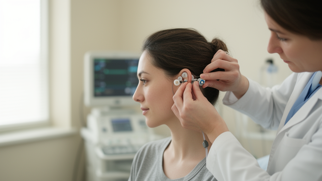
Electrical stimulation of nerves in the ear can prime human platelets and, in the case of vagus nerve stimulation, measurably speed up clot formation in the blood of healthy volunteers, according to a first-in-human exploratory trial published in Bioelectronic Medicine. The randomized, double-blind, sham-controlled study, registered as NCT05977946, tested two noninvasive neurostimulation approaches: transcutaneous auricular vagus nerve stimulation, or taVNS, which targets the auricular branch of the vagus nerve, and transcutaneous auricular neurostimulation, or tAN, which combines stimulation of that vagal branch with the auriculotemporal nerve, a sensory branch of the trigeminal nerve. Twenty-four healthy adults completed all phases of the study, and the stimulation was well tolerated, with no observable adverse events attributed to the devices during the study period.
Implanted cervical vagus nerve stimulation has treated medically refractory epilepsy and depression since 1997, and has more recently received regulatory approval for chronic ischemic stroke and rheumatoid arthritis. In preclinical hemorrhage models, invasive VNS accelerated clot initiation, increased clot deposition at injury sites, and reduced blood loss after peripheral soft tissue trauma in swine. Mouse studies traced the mechanism to acetylcholine-producing T lymphocytes in the spleen that contact circulating platelets: cholinergic signaling through platelet α7 nicotinic acetylcholine receptors raises intracellular calcium, which facilitates the secretion of prothrombotic α granules. This process, known as platelet priming, leaves platelets poised to form clots faster after tissue injury without necessarily triggering spontaneous clotting. Trigeminal nerve stimulation also reduced traumatic hemorrhage in mice, though its mechanism remains unknown, and a separate trial recently reported that tAN reduced blood loss and days of menstruation in women with heavy menstrual bleeding related to idiopathic or von Willebrand disease.
What remained unknown was whether these effects, demonstrated extensively in animals, could be reproduced with noninvasive ear stimulation in humans, and whether they would show up in standard laboratory measures of hemostasis. The research team, based at the Feinstein Institutes for Medical Research at Northwell Health, enrolled adults aged 18 to 65 while excluding anyone with a history of bleeding disorders, thrombocytopenia, anemia, thrombotic events such as stroke or pulmonary embolism, use of coagulation- or platelet-modifying therapies, recent nicotine use, or pregnancy. Twenty-five subjects were assessed for eligibility between November 2023 and August 2024; 24 met the criteria and were randomized in a 1:1 ratio, with 11 assigned to the tAN arm and 13 to the taVNS arm. Recruitment was halted after the first 24 subjects because of a performance site operational issue that precluded timely study completion.
The experimental design was deliberately conservative. Each participant first underwent a baseline blood draw, then received 30 minutes of sham stimulation through an identical earpiece device, followed by a post-sham blood draw ten minutes later. Only then did they receive 30 minutes of active stimulation, with blood drawn at ten minutes, one hour, and two hours afterward. Participants were blinded to both their group assignment and the timing of active versus sham stimulation, and were told they might or might not perceive any sensation. The research coordinator, who applied the assigned stimulation, was unblinded but did not discuss allocations with the study team. taVNS delivered pulses at 250 microseconds and 30 hertz to the vagal branch region, while tAN added 100 hertz stimulation to the auriculotemporal nerve region. Stimulation intensity started at 2.5 milliamperes and was reduced in small increments if participants reported discomfort.
The results split cleanly by modality. After taVNS, platelets exposed ex vivo to collagen showed significantly higher P-selectin expression, a marker of α granule secretion, ten minutes after stimulation compared with the post-sham baseline, rising from 2.97 to 4.55 with a p value of 0.03. After tAN, platelets exposed to ADP showed significantly higher expression of the activated GPIIb/IIIa fibrinogen receptor one hour after stimulation, rising from 19.7 to 25.79 with a p value of 0.035. In other words, the two stimulation types primed platelets through different signaling pathways: taVNS through collagen-mediated α granule release, and tAN through ADP-mediated conformational change of the primary fibrinogen receptor. Notably, thrombin exposure produced no changes in α granule secretion after either stimulation, a contrast with the murine data that the authors attribute to known differences in protease-activated receptor expression and signaling between mouse and human platelets.
The thromboelastography findings were equally striking and equally selective. In blood collected ten minutes after stimulation, taVNS significantly shortened the R time, which quantifies the speed of clot initiation, from 5.74 to 4.91 minutes with a p value of 0.004, a roughly 15 percent acceleration. It also shortened the K time, a measure of clot propagation speed, from 1.189 to 1.10 with a p value of 0.05, and increased the α angle, which reflects the rate of clot stabilization through fibrin cross-linking, from 73.78 to 75.53 degrees with a p value of 0.02. tAN, by contrast, changed none of these kinetic parameters. Neither stimulation altered maximum amplitude, a measure of overall clot strength, nor LY30, a measure of clot breakdown at 30 minutes, and circulating fibrinogen levels were unchanged. The authors suggest that taVNS may enhance fibrin cross-linking through a more favorable thrombotic environment on the surface of primed platelets rather than by raising fibrinogen availability.
Just as important were the parameters that did not move. Neither tAN nor taVNS changed white blood cell counts, hemoglobin, hematocrit, or platelet counts. Standard coagulation assays, including prothrombin time, partial thromboplastin time, international normalized ratio, and D-dimer, showed no differences compared with sham stimulation. Thrombin-antithrombin complex concentrations, measured both in circulating blood and in blood shed from a fingertip lancet injury as a model of local capillary bleeding, were also unchanged after either stimulation. Taken together, these null results suggest that at the prescribed stimulation settings the interventions did not push the coagulation cascades into systemic hypercoagulability, did not deplete or expand circulating cell populations, and did not amplify thrombin generation after superficial capillary injury. The authors note that finger lancets cause dermal capillary injury, whereas the preclinical models involved direct arterial or arteriolar disruption, which may explain the discrepancy with the animal thrombin findings.
Safety data were limited but reassuring within their scope. The single reported adverse event occurred during initial blood collection, when a participant experienced mild light-headedness that resolved without medical attention; the electrode had been applied but stimulation had not been activated, and the event was deemed unrelated to the intervention. Because adverse events were only assessed during the short study period, the authors caution that late-onset adverse events cannot be excluded. The trial also carried methodological limitations that the authors lay out explicitly. The cohort was mostly female, whereas most trauma patients are male. Sample sizes of 11 and 13 per group were small, and as an exploratory study it did not prespecify primary and secondary outcomes, which raises the risk of false positives across the many reported measures; the authors advise interpreting the p values with caution. Random TEG assay results from nine subjects, affected roughly equally across groups, fell outside the manufacturer’s recommended analysis window and were excluded, and two subjects received the incorrect stimulation paradigm but were retained in the intent-to-treat analysis. The unblinded coordinator role and the compressed sequence of sham and active stimulation with repeated blood draws within a single session add further caveats.
The authors are careful about what the findings do and do not demonstrate. Because all subjects were healthy and their thromboelastography results never left the normal range, it remains unclear whether the observed acceleration in clotting kinetics after taVNS would translate into improved clinical hemostasis, and no standardized method exists to predict clinical bleeding outcomes from the platelet priming assay used here. What the trial does establish is proof of mechanism: noninvasive ear stimulation can safely prime human platelets, and taVNS can accelerate clot initiation, propagation, and stabilization as quantified by viscoelastic testing. The authors argue that taVNS and tAN now warrant additional clinical study as potential therapies for traumatic or surgical hemorrhage and for congenital or acquired coagulopathies such as hemophilia A, hemophilia B, and von Willebrand disease, where preclinical VNS work showed hemostatic restoration despite factor VIII deficiency. They also speculate that combining these stimulations with tranexamic acid, which inhibits clot lysis, might prove synergistic in trauma-induced coagulopathy, and that higher intensities or longer stimulation durations might activate platelet responses capable of addressing more severe injuries. Larger clinical trials will be needed to determine whether faster clotting in a laboratory assay becomes less bleeding in the operating room or on the battlefield.
Subject of Research: Effects of transcutaneous auricular vagus nerve stimulation and combined vagal-trigeminal neurostimulation on platelet function and hemostasis in healthy adults
Article Title: Effects of transcutaneous auricular vagus nerve stimulation or combined vagal and trigeminal nerve stimulation on platelet function and laboratory hemostasis parameters in healthy human subjects
Article References: Huston, J. M., Bravo-Iñiguez, C. E., Papoin, J., Ahmad, M., Le, B., Mirro, I., McWade, M., Benner, C., Covalin, A., Czura, C. J., Khodaparast, N., & Blanc, L. (2026). Effects of transcutaneous auricular vagus nerve stimulation or combined vagal and trigeminal nerve stimulation on platelet function and laboratory hemostasis parameters in healthy human subjects. Bioelectronic Medicine, 12(1), Article 14. https://doi.org/10.1186/s42234-026-00208-w
Image Credits: AI Generated
DOI: 10.1186/s42234-026-00208-w
Keywords: transcutaneous auricular vagus nerve stimulation, taVNS, trigeminal nerve stimulation, platelet priming, thromboelastography, hemostasis, bioelectronic medicine, hemorrhage, coagulation, clinical trial, vagus nerve, blood clotting
News Source: Ophelia Keating. (October 29, 2026). Ear nerve stimulation primes platelets and speeds clotting in first-in-human trial. Scienmag.



