


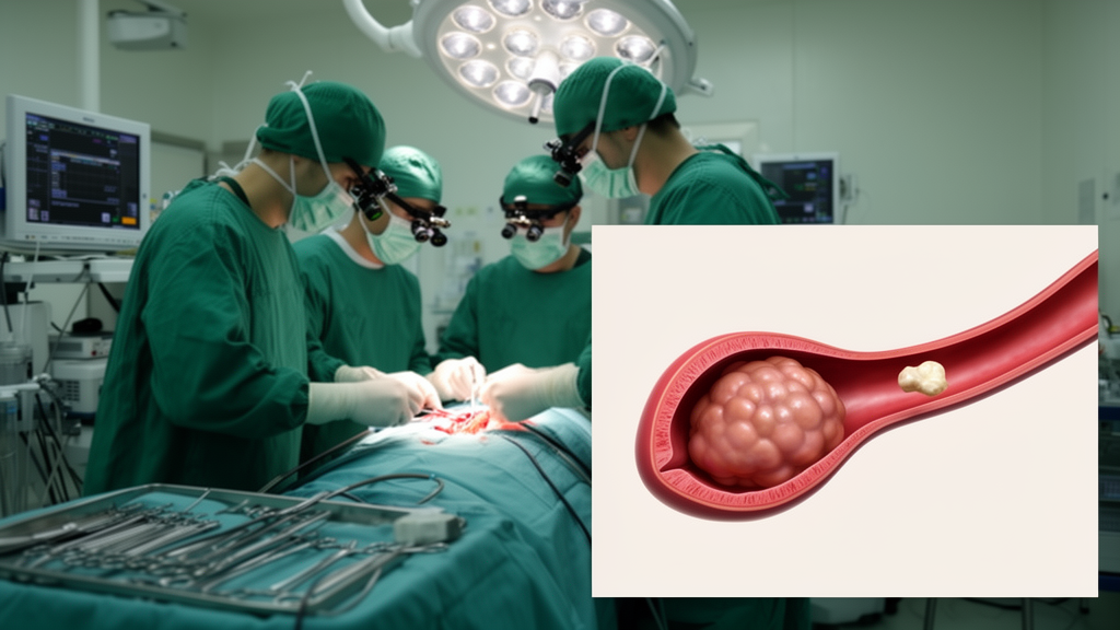
A 62-year-old woman arrived at a Japanese hospital with sudden weakness on her left side, loss of vision in the right half of her visual field, and a left arm that was cold to the touch. Imaging soon revealed the cause: a large tumor in her left atrium, the upper chamber of her heart, along with a blocked left subclavian artery and infarctions in her spleen and kidney. The tumor was a cardiac myxoma, the most common primary benign tumor of the heart, and the imaging also showed occlusion of the left subclavian artery and infarctions in her spleen and kidney. But what happened next turned the case from a routine tumor removal into a diagnostic puzzle that her surgical team has now documented in detail.
When surgeons prepared to remove the myxoma, repeated transthoracic echocardiography and abdominal ultrasound showed a 33-millimeter, immobile, linear hyperechoic structure in the suprarenal portion of the inferior vena cava, the large vein returning blood from the lower body to the heart. Color Doppler imaging demonstrated blood flow surrounding the structure in both long- and short-axis views, a pattern that suggested an organized thrombus lodged in the vein. The finding was consistent across scans performed on hospital days 18, 25 and 38, using different transducers, different examiners and multiple sessions. Abdominal ultrasonography showed the same structure. Yet contrast-enhanced computed tomography performed on admission and cardiac magnetic resonance imaging performed on hospital day 32 revealed no corresponding lesion in the inferior vena cava at all.
That discrepancy mattered enormously for surgical planning. Removing a cardiac myxoma requires cardiopulmonary bypass, which depends on cannulating the ascending aorta, the superior vena cava and the inferior vena cava. If a real thrombus sat in the inferior vena cava, standard cannulation through the right atrium could dislodge it and send it into the pulmonary circulation as a potentially fatal embolism. The team therefore faced a genuine dilemma: trust the repeated ultrasound findings and alter the operative approach, or trust the cross-sectional imaging and proceed conventionally.
The case, reported by Hiroto Yasumura and colleagues in the journal General Thoracic and Cardiovascular Surgery Cases, unfolds as a careful stepwise diagnostic workup. On arrival, laboratory tests had shown an elevated white blood cell count of 14,800 per microliter, decreased hemoglobin of 9.2 grams per deciliter, elevated C-reactive protein of 7.02 milligrams per deciliter, hyperfibrinogenemia of 624 milligrams per deciliter, and an elevated D-dimer of 4.7 micrograms per milliliter. Crucially, while C-reactive protein and fibrinogen remained elevated, subsequent D-dimer measurements returned to the normal range, which argued against active clot formation. Twelve-lead electrocardiography showed normal sinus rhythm with an enhanced second hump of the P wave in lead II but no enhanced negative deflection in lead V1, and chest radiography showed only minimal pulmonary congestion.
Transthoracic echocardiography on hospital day 2 had already characterized the cardiac tumor itself: a large, mobile, polypoid mass measuring 18 by 60 millimeters in the left atrium, its tail swinging back and forth between the left atrium and left ventricle in synchrony with ventricular contraction. The left atrium measured 32 millimeters, the diastolic left ventricle 45 millimeters, and the systolic left ventricle 32 millimeters, with a left ventricular ejection fraction of 55 percent. Valve regurgitation was trace at the aortic and tricuspid valves and mild at the mitral valve. Coronary computed tomography showed no coronary stenosis, and brain magnetic resonance imaging confirmed an acute infarction in the right occipital lobe, prompting oral apixaban from hospital day 4 to prevent further thromboembolic events.
Because hemorrhagic transformation of a cerebral infarction before cardiac surgery requiring cardiopulmonary bypass can be life-threatening, and because such transformation may occur within four weeks of an ischemic stroke, the team postponed the operation until hospital day 39. The authors also note that apixaban was chosen deliberately: compared with warfarin, apixaban has been associated with a significantly lower risk of major bleeding, particularly intracranial hemorrhage, while maintaining or improving protection against thromboembolic events. Since the anticoagulant was started on hospital day 4 and the inferior vena cava finding first appeared on hospital day 18, the team considered it unlikely that the drug contributed to the structure’s development.
On the day of surgery, the diagnostic picture shifted again. Preoperative transesophageal echocardiography performed under general anesthesia demonstrated no hyperechoic structure in the inferior vena cava in either long- or short-axis views. Hemodynamic assessment with a Swan Ganz catheter inserted via the right internal jugular vein showed a pulmonary artery pressure of 27/12 millimeters of mercury and a central venous pressure of 7 millimeters of mercury. Given the residual concern about a possible thrombus, the team took no chances: after median sternotomy, cardiopulmonary bypass was initiated via cannulation of the ascending aorta and superior vena cava, but the inferior vena cava cannula was inserted through the right femoral vein under fluoroscopic guidance, avoiding the right atrial route altogether.
What followed settled the question definitively. After cardiac arrest and right atriotomy, direct inspection of the suprarenal inferior vena cava and right atrium revealed neither thrombus nor any abnormal anatomical structure such as a Chiari network or a prominent Eustachian valve, the congenital variants that can mimic intraluminal lesions. The left atrial tumor, attached to the fossa ovalis, was excised en bloc through a right-sided left atriotomy. The patient was weaned from cardiopulmonary bypass without difficulty, histopathological examination confirmed the diagnosis of cardiac myxoma, and transthoracic echocardiography on postoperative day 7 showed complete disappearance of the inferior vena cava hyperechoic structure. She was discharged on postoperative day 22, and follow-up echocardiography on postoperative day 204 demonstrated no recurrence of either the myxoma or the venous finding.
So what had the ultrasound been seeing? The authors consider several mechanisms. One leading candidate is spontaneous echo contrast, a swirling smoke-like echogenic appearance associated with low-flow states and blood stasis, thought to arise from aggregation of red blood cells mediated by fibrinogen under low-flow conditions. Its predisposing factors include atrial fibrillation, structural cardiovascular abnormalities, aging, low blood flow velocity, reduced shear rate, an elevated erythrocyte sedimentation rate, hyperfibrinogenemia and high hematocrit. Previous case reports have described hyperechoic spontaneous echo contrast within the inferior vena cava, particularly in patients with constrictive pericarditis, where it closely mimicked thrombus. Notably, the patient in this case did have hyperfibrinogenemia, although the authors point out there was little clinical or hemodynamic evidence of significant venous stasis. Importantly, an earlier study of 106 consecutive adults evaluated for spontaneous echo contrast in the inferior vena cava found that such contrast may also appear in individuals without apparent pathology and may not be clinically significant.
Artifacts remain firmly on the differential list. Side-lobe and beam-width artifacts can produce spurious intraluminal echoes and false-positive findings, and the authors note that no previous report has specifically described an inferior vena cava hyperechoic structure caused by these echogenic artifacts. The mirror-image artifact offers another possibility: differences in the transit time of reflected ultrasound waves can create an inverted image of the inferior vena cava at a deeper location or on the opposite side of the true vessel, in which case the posterior wall of the true vessel between the true and false images may appear as a hyperechoic structure resembling a thrombus within a dilated vein. The authors acknowledge that an imaging-related pseudo-structure cannot be completely excluded in their case. Paradoxically, the very consistency of the finding, seen in both long- and short-axis views on repeated scans with different transducers and examiners across multiple days, makes a typical artifact less likely, yet direct intraoperative inspection failed to identify any intraluminal lesion. Taken together, the team concluded the structure was more likely a pseudo-thrombus than a true thrombus.
The broader lesson the authors draw is about integrating discrepant findings from multiple imaging modalities rather than relying on any single test. Transesophageal echocardiography has been reported to have a sensitivity of 68 percent and a specificity of 98 percent for detecting intracardiac thrombi, and a sensitivity of 97 percent and specificity of 99 percent for diagnosing aortic dissection, figures that underscore why no single modality should be treated as definitive. Based on their experience, the team suggests that when a similar inferior vena cava finding is encountered, additional transthoracic echocardiography in different patient positions and transesophageal echocardiography performed before induction of general anesthesia should be considered preoperatively, since these approaches may help distinguish a true thrombus from a pseudo-thrombus. They also note that contrast-enhanced cardiac magnetic resonance imaging, transesophageal echocardiography before induction of anesthesia, and transthoracic imaging in the lateral decubitus position might have provided additional information in their own case, as the patient was scanned in the supine position during the original echocardiography. To their knowledge, an inferior vena cava hyperechoic structure associated with cardiac myxoma had not been previously reported, making this a rare and instructive addition to the literature on a diagnostic pitfall that could, in other hands, have changed the course of a cardiac operation.
Subject of Research: A surgical case report of an inferior vena cava hyperechoic structure mimicking thrombus in a patient with cardiac myxoma
Article Title: Inferior vena cava hyperechoic structure in a patient with cardiac myxoma: a surgical case report and literature review
Article References: Yasumura, H., Matsuba, T., Ibi, R., Minagoe, S., Hiramine, A., Ogata, Y., Shimoishi, K., Fukumoto, Y., Yotsumoto, G., & Soga, Y. (2026). Inferior vena cava hyperechoic structure in a patient with cardiac myxoma: a surgical case report and literature review. General Thoracic and Cardiovascular Surgery Cases, 5(1), Article 42. https://doi.org/10.1186/s44215-026-00277-y
Image Credits: AI Generated
DOI: 10.1186/s44215-026-00277-y
Keywords: cardiac myxoma, inferior vena cava, echocardiography, pseudo-thrombus, spontaneous echo contrast, case report, cardiopulmonary bypass, transesophageal echocardiography, ultrasound artifacts, multimodality imaging, thromboembolism, cardiac surgery
News Source: Ophelia Keating. (October 29, 2026). Research report: Inferior vena cava hyperechoic structure in a patient with cardiac myxoma: a surgical case report and literature review. Scienmag.



