A 63-year-old man with a rare pineal region tumor is reported to have achieved a favorable neurological recovery after a staged treatment plan that combined cerebrospinal fluid diversion, near-total surgical excision and focal radiotherapy without chemotherapy. The tumor was a pineal parenchymal tumor of intermediate differentiation, or PPTID, classified as CNS WHO grade 3, and the case is described in the Egyptian Journal of Neurosurgery as a case report with a contemporary literature review. The authors report that a telephone review ten months after surgery and external beam radiotherapy found the patient conscious and oriented, with occasional headache, no diplopia, a Karnofsky performance status score of 90, and allegedly no residual lesion on post-radiotherapy MRI.
PPTIDs are uncommon neoplasms that represent a small subset of pineal region tumors. Their rarity, variable biological behavior and evolving pathological classification make management challenging, particularly in the absence of standardized treatment guidelines. Within the WHO classification, PPTID can be graded as WHO grade 2 or 3, a categorization that was introduced in the 2007 classification and remains unchanged in the 2021 classification, positioning the tumor as an intermediate malignancy. The authors note that PPTIDs account for 13 to 20 percent of all pineal parenchymal tumors, with variation across several literature reviews. Pineal region tumors themselves are rare, comprising less than 1 percent of central nervous system tumors, and their deep-seated location, proximity to the great cerebral and basal veins, difficult histopathological differentiation and lack of standardized treatment protocols add therapeutic complexity.
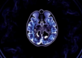
The patient presented with intermittent holocranial headache of one month’s duration that was relieved with vomiting. On examination, his Glasgow Coma Scale score was 15 and he had minimal upward gaze palsy with no gross neurological deficits. His pupils were bilaterally equal and reactive to light, and Romberg’s test and cerebellar signs were negative. He had previously undergone endoscopic third ventriculostomy after being diagnosed with a pineal region lesion with obstructive hydrocephalus, and he had no significant comorbidities. Contrast MRI of the brain showed a well-defined, heterogeneously post-contrast enhancing, T2 hyperintense lobulated lesion in the pineal region with minimal internal bleed. The lesion was located superior to the tectal plate with mass effect, and the current scan showed no hydrocephalus.
Definitive surgery was performed through a midline suboccipital craniotomy with a supracerebellar infratentorial approach, also known as the Krause approach. The patient was positioned prone with the head fixed in a Mayfield clamp. After a midline skin incision from 3 centimeters above the inion to the C2 spinous process, a suboccipital craniotomy was carried out with two burr holes below the transverse sinus, exposing the margin of the transverse sinus bilaterally. The dura was opened in a U-shaped fashion and tented cranially, and minor bridging veins were coagulated and divided. Thickened arachnoid was noted overlying the pineal lesion and was sharply divided. The vein of Galen and the precentral cerebellar vein were identified and safeguarded, and the tumor, which lay deeper to the precentral cerebellar vein, was reached after that vein was coagulated and divided.
The lesion was described as pinkish, soft and highly vascular, with good planes all around, and near-total decompression was achieved. The thalamus and third ventricle were visualized at the end of resection, and a gelatin sponge was placed in the third ventricle to prevent intraventricular hemorrhage. A frozen section sample was reported as an intermediate grade pineal tumor. The authors state that complete tumor excision was not attempted because of the close relationship of the lesion with critical neurovascular structures in the pineal region, where aggressive resection could have led to significant neurological deficits. Fascia lata harvested from the left thigh was used for a lax duraplasty, the bone flap was replaced with titanium plates and screws, and multilayered closure was completed.
Postoperatively, the patient was extubated on the table and shifted to the intensive care unit. Contrast CT of the brain the next day showed complete excision of the lesion with minimal pneumocephalus and no hydrocephalus. He had minimal upward gaze palsy with convergence retraction nystagmus after the operation, which gradually subsided. Mechanical DVT prophylaxis was strictly followed during the hospital stay, he was gradually mobilized with support, and he improved clinically and was discharged in stable condition.
The histopathological workup was central to establishing the diagnosis. On light microscopy, the tumor was composed of diffuse sheets of tumor cells displaying a lobulated pattern of arrangement, intersected by delicate fibrovascular septae and a loose fibrillary matrix. The tumor cells had speckled nuclear chromatin, inconspicuous nucleoli and scant eosinophilic cytoplasm. Occasional pseudorosettes, mitotic activity of 5 to 6 per ten high-power fields and mild nuclear pleomorphism were noted. Immunohistochemistry showed positivity for synaptophysin, CD56 and vimentin, and negativity for CD99, NeuN and OLIG2. INI-1 was retained and the Ki-67 proliferation index was 30 percent. The negative OLIG2 result, a marker of glial differentiation, made a glioma unlikely, and the histomorphology was not consistent with a germ cell tumor, with negative PLAP immunostaining effectively ruling out germinoma. Synaptophysin positivity together with round cell morphology and finely speckled salt-and-pepper chromatin favored PPTID, and the increased mitotic activity and high Ki-67 index led to categorization as CNS WHO grade 3.
After multidisciplinary tumor board discussion, the team decided to give local radiation therapy to the resection cavity alone without chemotherapy. The authors explain that adjuvant therapy was limited to local radiotherapy because there was no evidence of tumor dissemination, and that craniospinal irradiation was not considered given the absence of radiological or clinical evidence of cerebrospinal fluid spread and the potential for unnecessary treatment-related morbidity. Chemotherapy was not administered because its role in localized PPTID remains unclear. The patient’s age, good postoperative neurological status, absence of leptomeningeal spread and near-total excision influenced the decision. Observation alone was deemed inadequate given the high mitotic index and proliferative activity, so a balanced approach combining surgery and focal radiotherapy was adopted to maximize disease control while preserving quality of life. External beam radiotherapy was delivered in 30 fractions.
The discussion places the case in a wider clinical context. Based on a review of 72 cases, the commonest presentation was noted to be headache, followed by diplopia, vertigo, paraparesis and unstable gait, and Parinaud syndrome is reported as common in 75 percent of all pineal tumors, particularly in the pediatric population. This patient presented with isolated holocranial headache without Parinaud syndrome. Radiologically, PPTIDs on CT usually appear as well-defined iso- to hyperdense pineal region masses with variable calcification and contrast enhancement, while MRI typically demonstrates a well-lobulated lesion that is T1 iso- to hypointense and heterogeneously T2 hyperintense with moderate to marked post-contrast enhancement. Compared with pineoblastomas, PPTIDs tend to be more circumscribed and exhibit more indolent behavior, though overlap with germ cell tumors is common, so histopathological confirmation remains mandatory. The imaging characteristics in this patient were consistent with the previously described classical features of PPTID.
On management principles, the authors note that because of the rarity of the disease, treatment strategies are largely based on retrospective series. Maximal safe surgical resection is universally accepted as the primary treatment modality in CNS tumors, providing both diagnostic tissue and local disease control, and several studies have demonstrated improved outcomes following gross or near-total resection. Adjuvant treatment is tailored according to tumor grade, extent of resection and evidence of dissemination, with surgery alone potentially sufficient for selected grade 2 tumors, while grade 3 lesions generally warrant adjuvant radiotherapy, and craniospinal irradiation is reserved for cases with cerebrospinal fluid dissemination. In this case, early cerebrospinal fluid diversion via endoscopic third ventriculostomy relieved obstructive hydrocephalus and stabilized intracranial pressure before definitive surgery, facilitating safe resection and optimizing postoperative recovery. On molecular pathology, the authors note that KBTBD4 in-frame insertions are a desirable criterion for PPTID diagnosis and have emerged as a useful molecular marker supporting the diagnosis. Given the rarity of PPTID and the lack of uniform treatment guidelines, the authors conclude that future studies with larger patient cohorts and long-term follow-up are needed to better define the role of adjuvant therapies, that future multicentric studies integrating molecular profiling with clinical outcomes are required to refine risk stratification and establish standardized protocols, and that long-term follow-up is crucial due to the risk of recurrence.
Subject of Research: A case report of a CNS WHO grade 3 pineal parenchymal tumor of intermediate differentiation treated with staged endoscopic third ventriculostomy, near-total resection and focal radiotherapy.
Article Title: Parenchymal tumor of the pineal region: insights from a rare case and contemporary literature review
Article References: Ravichandran, P., Kapilavayi, S., K R, S. B., Balasundaram, P., & Sundaravadivelu, V. (2026). Parenchymal tumor of the pineal region: insights from a rare case and contemporary literature review. Egyptian Journal of Neurosurgery, 41(1), Article 174. https://doi.org/10.1186/s41984-026-00669-w
Image Credits: AI Generated
DOI: 10.1186/s41984-026-00669-w
Keywords: pineal parenchymal tumor of intermediate differentiation, PPTID, CNS WHO grade 3, pineal region tumor, endoscopic third ventriculostomy, supracerebellar infratentorial approach, near-total resection, focal radiotherapy, KBTBD4, case report, Ki-67 index, multidisciplinary tumor board
News Source: Nathaniel Bowman. (October 28, 2026). Rare Pineal Region Tumor Managed with Staged Surgery and Focal Radiotherapy in 63-Year-Old Man. Scienmag.