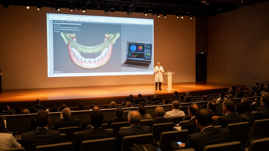
Bauru, Brazil — The VIII International Symposium on Orofacial Clefts and Related Anomalies, held at the Bauru Medical School of the University of São Paulo (FMBRU-USP), drew together clinicians, researchers, students, and healthcare professionals for three days of discussion on the latest advances in craniofacial rehabilitation. Organized by the university’s Graduate Program in Rehabilitation Sciences, the meeting ran alongside the first Pre-Symposium of the graduate program and featured a keynote lecture on Robin Sequence management protocols, six thematic panels, and a poster session showcasing 84 accepted presentations drawn from 90 submitted abstracts.
The symposium carried particular institutional weight this year. Following the incorporation of the Hospital for Rehabilitation of Craniofacial Anomalies (HRAC-USP) into the Bauru Clinical Hospital, the graduate program was transferred in 2025 to the Bauru Medical School. In the same year, the program achieved a grade 6 out of 7 from CAPES, the Brazilian federal agency responsible for regulating and funding graduate education, a rating that recognizes scientific productivity and internationalization. To mark this transition, the organizing committee introduced the First Pre-Symposium, which showcased the newly established research area of Interdisciplinary Care Across the Life Cycles and allowed faculty advisors to present their research lines while integrating newly admitted master’s and PhD students into the program before the main scientific meeting began.
The keynote lecture that opened the scientific program was delivered by Professor Christian Friedrich Poets, a specialist in neonatology and sleep medicine who heads the Department of Neonatology at University Hospital Tübingen in Germany. He presented the Tübingen Palatal Plate, a device developed by his team to treat upper airway obstruction in infants with Robin Sequence. The appliance consists of a palatal plate fitted with an individualized velar extension that repositions the tongue base, thereby relieving airway obstruction while avoiding more invasive interventions such as tracheostomy or mandibular distraction osteogenesis. Professor Poets presented highly favorable long-term outcomes from the Tübingen experience, and stated that the protocol has since been adopted by several centers worldwide. He cautioned, however, that the device requires careful customization and specialized follow-up adjustments because of the complexity of upper airway anatomy, particularly in infants with syndromic Robin Sequence.
A second keynote from Professor Marielena Layuno-Matos, a craniofacial orthodontist affiliated with the University of Puerto Rico who completed specialized training at Seattle Children’s Hospital, described how that institution has adapted a similar approach. Initial management there consists of conservative measures including side- and prone-positioning, nasogastric feeding, and respiratory support. Historically, patients who required escalation in care were considered for surgical intervention such as mandibular distraction osteogenesis or tracheostomy. In August 2023, Seattle Children’s introduced the Orthodontic Airway Plate, an appliance inspired by the Tübingen Palatal Plate that displaces the base of the tongue forward and maintains upper airway patency. The institution’s initial clinical experience was presented through case examples, emphasizing interdisciplinary workflows involving otolaryngology, craniofacial orthodontics, feeding specialists, bioengineering, developmental therapists, and nursing.
The first thematic panel, dedicated to Robin Sequence treatment, compared the multidisciplinary protocols adopted by rehabilitation centers in Tübingen, Seattle, and Bauru. Panelists discussed both intraoral devices, which relieve upper airway obstruction by repositioning the tongue base, and the reality that limited availability of these appliances means conservative alternatives remain essential. Non-invasive techniques such as the prone position provide immediate but not long-lasting relief of respiratory distress, while the nasopharyngeal cannula offers greater effectiveness than prone positioning alone. Professor Sergio Henrique Kiemle Trindade presented how treatment can be guided by objective assessment through polysomnography to quantify airway obstruction and monitor outcomes. When conservative approaches fail or severe obstruction is present, surgical management becomes necessary. Professor Cristiano Tonello highlighted mandibular distraction osteogenesis as the preferred surgical option because it provides rapid improvement in both airway patency and feeding. Glossopexy, by contrast, is currently less favored and should be considered only after careful multidisciplinary discussion, while tracheostomy remains an option for the most severe cases when other interventions are unsuccessful.
The panel also stressed the importance of long-term follow-up after surgical interventions and of routine genetic evaluation, because approximately 50 percent of Robin Sequence cases are syndromic, meaning they are associated with other anomalies. Distinguishing between isolated and syndromic forms is crucial for determining the steps of the rehabilitation process, since the interventions may differ and accurate genetic counseling depends on that distinction. The panelists concluded that Robin Sequence demands individualized, multidisciplinary management based on the severity of upper airway obstruction, with the palatal plate devices emerging as an innovative, minimally invasive option for selected patients and objective airway assessment remaining essential to optimizing clinical outcomes.
Digital technologies and artificial intelligence formed the focus of the second panel, which examined how these tools have enhanced pediatric dental care for patients with cleft lip and palate by supporting clinical documentation, objective assessment, and interdisciplinary treatment planning. Recent advances include digital intraoral scanning, three-dimensional assessment of dental arches and craniofacial growth, and stereophotogrammetry for longitudinal facial analysis. As craniofacial growth approaches completion, orthognathic surgery becomes an essential component of comprehensive rehabilitation for patients presenting residual skeletal discrepancies, requiring individualized interdisciplinary assessment. The panel also discussed early orthognathic intervention in selected adolescents, weighing potential functional, esthetic, and psychosocial benefits against the risks associated with residual craniofacial growth, relapse, and the potential need for retreatment. Complementing these advances, artificial intelligence is increasingly supporting diagnosis, treatment planning, and outcome assessment in cleft care, with AI-based approaches developed for automated cleft morphology and occlusion classification as well as three-dimensional image segmentation, reconstruction, and objective evaluation of alveolar bone graft outcomes, carrying the potential to improve diagnostic accuracy, prediction of treatment outcomes, and the reproducibility of quantitative analyses.
The pressure-flow technique was presented as an objective method for assessing velopharyngeal function by indirectly estimating the velopharyngeal orifice area during speech production, providing quantitative measurements that support clinical decision-making. Surgical management at HRAC-USP is guided by perceptual-auditory and instrumental assessments together with the size of the velopharyngeal gap, closure pattern, and the mobility of the soft palate and pharyngeal walls. Treatment is individualized according to velar anatomy and function, gap size, and lateral pharyngeal wall movement, assessed mainly by nasopharyngoscopy and videofluoroscopy. Palate re-repair with radical intravelar veloplasty is preferred when levator abnormalities or velar asymmetry are present. For persistent insufficiency with adequate palatal function, posterior pharyngeal wall augmentation may be used for small gaps, while a superiorly based pharyngeal flap is indicated for larger gaps with adequate lateral wall movement. For patients who are not candidates for surgery, rehabilitation may be achieved with a palatal prosthesis incorporating a pharyngeal bulb, developed through collaboration between speech-language pathologists and dental professionals, combined with targeted speech therapy.
Evidence from long-term clinical research anchored the fourth panel on rehabilitation outcomes. Panelists highlighted the contribution of the Timing of Primary Surgery for Cleft Palate trial, which demonstrated that children who underwent palatoplasty at 6 months of age exhibited a higher frequency of canonical babbling at 12 months and a lower prevalence of velopharyngeal insufficiency at 5 years in the primary analysis, while no clinically meaningful differences were observed between the groups in dentofacial outcomes. The Florida Project, developed at HRAC-USP, evaluated institutional outcomes while incorporating training in international surgical techniques, and its findings showed that techniques promoting more effective muscle reconstruction achieve excellent speech and velopharyngeal function, although potential effects on facial growth must also be considered. As these cohorts have now reached adulthood, long-term functional, structural, and aesthetic outcomes can be comprehensively assessed. The panel also highlighted the increasing role of stereophotogrammetry as a non-invasive, three-dimensional technology for objectively evaluating facial morphology, comparing surgical protocols, and monitoring craniofacial growth over time, reducing the subjectivity of conventional facial analysis.
The final panels broadened the scope to translational research and international collaboration. Panelists presented advances in the neurophysiology of hearing, the application of proteomics as an emerging strategy for investigating craniofacial anomalies through protein profiling that captures the combined effects of genetic and environmental factors, and the transformation of molecular genetics through next-generation sequencing methodologies that enable more comprehensive characterization of the genetic basis of these conditions. The integration of multi-omics data with detailed phenotypic characterization was emphasized as essential for improving understanding of genotype–phenotype relationships, with conditions such as craniofacial microsomia illustrating the heterogeneous clinical manifestations and genetic variants involved. Computed tomography with three-dimensional reconstruction and magnetic resonance imaging were highlighted for their complementary roles in evaluating syndromic craniosynostoses, where comprehensive characterization of craniofacial and intracranial abnormalities supports individualized treatment planning. A closing panel on internationalization addressed how academic mobility, institutional partnerships, researcher exchange, multicenter collaborations, and research funding strengthen research capacity, standardize clinical protocols, and accelerate the translation of scientific discoveries into clinical practice. The symposium, which has accumulated more than 500 abstract submissions and drawn over 1,000 attendees across its successive editions, brought together 200 participants this year. This proceedings paper summarizes the principal discussions and key messages presented during the meeting, providing an overview of current advances, persistent challenges, and future directions in research, education, and the multidisciplinary management of individuals with orofacial clefts and related craniofacial anomalies.
Subject of Research: Advances in the multidisciplinary management of orofacial clefts and related craniofacial anomalies, including Robin Sequence airway devices, digital and AI technologies, velopharyngeal insufficiency treatment, and translational research.
Article Title: Proceedings from the VIII International Symposium on Orofacial Clefts and Related Anomalies – Bauru Medical School, University of São Paulo (FMBRU-USP)
Article References: Braga, B. M. R., Segarra, V. C. D., Miranda, A. F. D., Nogueira, A. C. P. A.-Y., Minozzi, A. C. C., Farha, A. L. H., de Oliveira Domingues, C. S. V., Queiroz, D. S. R., da Silva Nascimento, F., Botari, G. B., Baessa-Cardoso, G., Serigatto, H. R., Neves, M. C., Batista, N. T., Ferreira, R. A., da Silva, T. A., de Abreu Francisco, V., Layuno Matos, M., Poets, C. F., … Trindade, S. H. K. (2026). Proceedings from the VIII International Symposium on Orofacial Clefts and Related Anomalies – Bauru Medical School, University of São Paulo (FMBRU-USP). BMC Proceedings, 20(S35), Article 38. https://doi.org/10.1186/s12919-026-00401-4
Image Credits: AI Generated
DOI: 10.1186/s12919-026-00401-4
Keywords: orofacial clefts, Robin Sequence, Tübingen Palatal Plate, Orthodontic Airway Plate, velopharyngeal insufficiency, artificial intelligence, stereophotogrammetry, craniofacial rehabilitation, next-generation sequencing, TOPS trial, Bauru Medical School, multidisciplinary care
News Source: Drew Townsend. (October 28, 2026). Robin Sequence Airway Plates and AI Tools Take Center Stage at Brazilian Craniofacial Symposium. Scienmag.