


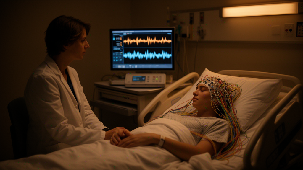
Seizures have been the most common neurological symptom in children with COVID-19, and they often occur without fever, distinguishing them from ordinary febrile seizures in their age of onset. A new study published in Brain Informatics set out to determine whether electroencephalography, or EEG, can reveal distinct brain network signatures that separate seizures provoked by COVID-19 from febrile seizures and from healthy brain activity.
Researchers collected EEG data from three groups: patients with febrile seizures unrelated to COVID-19, a group designated FS-N; patients with seizures provoked by COVID-19, designated PSP-COV; and healthy controls, designated HC. From these recordings they constructed causal brain networks using a technique called conditional Granger causality, or CGC, which estimates directed influences between signals recorded at different brain regions.
Once the networks were built, the team focused on causal connections that showed statistically significant group differences. These differing connections served two purposes: they were used as features for machine learning classification, and they helped identify the brain areas most affected in each condition.
A key finding concerned sleep. The CGC connections showed more significant group differences when measured in the sleep state than in the awake state, pointing to a sleep-specific disruption of EEG causal networks in seizures provoked by COVID-19.
The machine learning results were strong across all comparisons. When connections with group differences were used as classification features, accuracy reached 92.86 percent between FS-N and healthy controls, 90.9 percent between PSP-COV and healthy controls, and 84.62 percent between PSP-COV and FS-N. The three most discriminative connections separating provoked seizures from febrile seizures were found during sleep.
Examining which connections contributed most to classification, the researchers found that high-contribution connections featured positive out-in degrees in the frontal and temporal lobes, whereas the parietal and occipital lobes showed negative values. In other words, frontal and temporal regions acted predominantly as sources of causal influence, while parietal and occipital regions acted as targets.
One connection pattern stood out in the comparison between the two seizure groups: notably altered connections running from the posterior temporal lobe to the parietal lobe. This directed shift, from temporal sources toward parietal targets, offers a candidate neuroimaging biomarker for distinguishing COVID-19 provoked seizures from febrile seizures.
The authors suggest that these features, with frontal and temporal lobes as sources and parietal and occipital lobes as targets in the EEG causal network, offer new insights into the brain network mechanisms underlying seizures provoked by COVID-19. Because the study is based on EEG recordings from the collected patient and control groups, the reported accuracies and network patterns describe the measured cohorts, and the findings are presented as observational network features rather than established clinical diagnostic criteria.
Subject of Research: EEG-based causal brain network analysis of seizures provoked by COVID-19 compared with febrile seizures and healthy controls
Article Title: The brain causal network features in seizures provoked by COVID-19 using EEG
Article References: Zhang, Q., Li, S., Wu, L., Deng, C., Zhang, J., Wang, J., Cui, Q., Hu, W., Ju, R., Chen, H., & Gao, Q. (2026). The brain causal network features in seizures provoked by COVID-19 using EEG. Brain Informatics. https://doi.org/10.1186/s40708-026-00339-5
Image Credits: AI Generated
DOI: 10.1186/s40708-026-00339-5
Keywords: EEG, COVID-19, seizures, febrile seizures, causal brain networks, Granger causality, machine learning, frontal lobe, temporal lobe, parietal lobe, sleep, Brain Informatics
News Source: Cassandra Pierce. (October 28, 2026). EEG Causal Networks Reveal Sleep-Specific Brain Disruption in COVID-19 Provoked Seizures. Scienmag.



