Researchers report that a specific set of focused ion beam (FIB) milling settings can produce cross sections of proton exchange membrane fuel cell (PEMFC) cathode catalyst layers that are suitable for consistent two-dimensional pore-area analysis, without the need for cryogenic sample preparation. The study, published in Applied Microscopy, systematically examined how acceleration voltage, beam current, and temperature influence the quality of cross sections prepared for scanning electron microscopy.
In the work, Ji Hee Kwon, Jinnyeong Jo, and Byeong-Seon An used focused ion beam–scanning electron microscopy (FIB–SEM) to prepare and image cross sections of the catalyst layer under a range of conditions. Cross sections were milled at 5 kV and at 30 kV at room temperature, and at 30 kV under cryogenic conditions at −150 °C, allowing direct comparison of the resulting pore morphology across milling parameters.
The key finding concerns artifact control. According to the abstract, milling at 30 kV and 0.26 nA effectively minimized these curtaining artifacts. Under this condition, the apparent cross-sectional pore morphology was similar to that obtained by cryogenic milling at 30 kV and −150 °C.
The authors indicate that optimized room-temperature milling can provide cross sections suitable for consistent two-dimensional pore-area analysis under the conditions examined.
The reported result is deliberately scoped. The conclusion applies to the acceleration voltages, beam currents, and temperature conditions tested in the study, and the finding concerns cross-sectional preparation and two-dimensional image-based pore analysis rather than measured porosity values of specific commercial catalyst layers or fuel cell performance outcomes. Representative images in the paper show the range of milling conditions, with the optimized room-temperature condition of 30 kV and 0.26 nA highlighted for direct comparison against the corresponding cryogenic condition. For laboratories analyzing PEMFC electrode microstructure with FIB–SEM, the study offers a practical parameter set for producing artifact-minimized cross sections at room temperature, though the authors frame the benefit as consistent image-based analysis under the examined conditions rather than a universal prescription for all catalyst-layer materials or imaging systems.
Subject of Research: Optimization of focused ion beam milling parameters for preparing artifact-minimized cross sections of PEMFC cathode catalyst layers for porosity analysis.
Article Title: Imaging optimization of FIB cross sections for reliable porosity analysis of PEMFC cathode catalyst layers
Article References: Kwon, J. H., Jo, J., & An, B.-S. (2026). Imaging optimization of FIB cross sections for reliable porosity analysis of PEMFC cathode catalyst layers. Applied Microscopy, 56(1), Article 23. https://doi.org/10.1186/s42649-026-00142-w
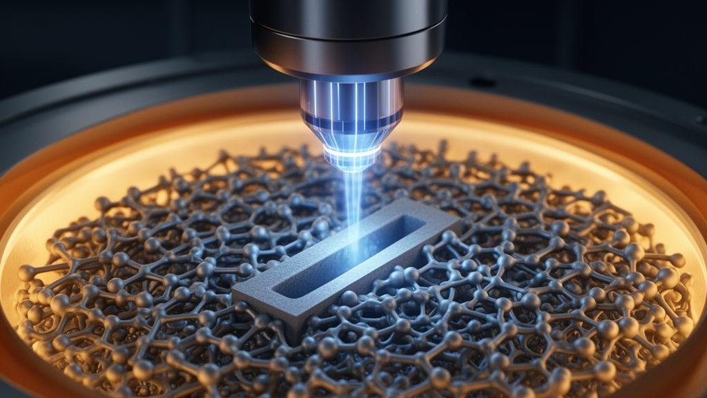
Image Credits: AI Generated
DOI: 10.1186/s42649-026-00142-w
Keywords: FIB-SEM, PEMFC, cathode catalyst layer, porosity, curtaining artifacts, cryogenic milling, cross-section imaging, fuel cells, electron microscopy, sample preparation, Imaging, optimization
News Source: Bethany Barker. (October 28, 2026). Optimized FIB milling yields reliable pore images of fuel cell catalyst layers. Scienmag.