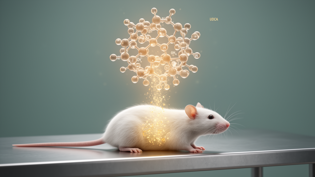
Ursodeoxycholic acid, a bile acid already approved for treating primary biliary cholangitis, lowered blood glucose and eased several metabolic disturbances in a rat model of type 2 diabetes, according to a study published on 31 July 2026 in Discover Endocrinology and Metabolism.
The research team set out because bile acids are known to do far more than digest fat. Beyond their traditional roles in fat absorption and cholesterol balance, bile acids act as signaling molecules that bind receptors such as the farnesoid X-activated receptor (FXR) and Takeda G-protein-coupled receptor-5 (TGR5). Activation of these receptors participates in the regulation of glucose and lipid metabolism, insulin sensitivity and energy homeostasis. Because type 2 diabetes is characterized by disordered glucose and lipid metabolism, targeting the bile acid signaling pathway has been proposed as a potential therapeutic strategy alongside conventional glucose-lowering drugs. Yet the efficacy of ursodeoxycholic acid in type 2 diabetes remained unclear, and previous studies have reported conflicting results.
To test the question, the researchers worked with 30 male Sprague-Dawley rats obtained from the Experimental Animal Center of Shanxi Medical University in Taiyuan, China. After a week of acclimatization, the animals were randomly divided into three groups of ten using a random number table. A normal control group received a standard diet containing 66.5% carbohydrate, 10.2% fat and 23.3% protein. The two remaining groups were fed a high-fat diet with 38.0% carbohydrate, 54.0% fat and 7.0% protein for four weeks. At the end of that fourth weekend, both high-fat groups received a single intraperitoneal injection of streptozotocin at 30 milligrams per kilogram, dissolved in 0.05 M citrate buffer at pH 4.4.
One of the diabetic groups then received daily oral gavage of ursodeoxycholic acid at a dose of 0.25 grams per kilogram, dissolved in normal saline, for four weeks. The diabetic control group and the normal control group received equal volumes of saline by gavage over the same period. All animals had free access to food and water, and the procedures were approved by the Institutional Animal Care and Use Committee of the Second Hospital of Shanxi Medical University. At the end of the experiment, the researchers performed oral glucose tolerance tests, measured serum biochemistry, insulin and C-peptide levels, quantified liver triglyceride and total cholesterol content, examined pancreatic tissue under the microscope, and used real-time PCR to measure the expression of nine genes involved in bile acid, lipid and glucose metabolism in the liver.
The model worked as intended. Four weeks after the streptozotocin injection, the diabetic rats showed significantly higher fasting blood glucose and higher one-hour and two-hour postprandial glucose than the normal controls, with all comparisons reaching a p-value below 0.001. Their body weights and serum levels of the liver enzymes ALT, ALP and GGT, along with serum triglycerides and total cholesterol, were also significantly elevated. Using the study’s diagnostic threshold of a fasting blood glucose of at least 7.24 millimoles per liter, the diabetic group met the criteria the authors used to identify type 2 diabetes rats, confirming that the model had been successfully established.
Ursodeoxycholic acid treatment changed the picture. Compared with the untreated diabetic rats, the treated animals showed significantly lower fasting blood glucose (p = 0.005) and lower two-hour postprandial glucose (p = 0.041). The area under the glucose curve during the oral glucose tolerance test decreased with treatment (p = 0.021), while the areas under the insulin curves increased (p = 0.047) and fasting insulin levels rose significantly (p = 0.019). Fasting C-peptide levels, a marker of endogenous insulin production, also increased with treatment (p = 0.002). Beta cell function, assessed by the homeostasis model, improved from 42.71 plus or minus 4.86 to 58.76 plus or minus 7.80 mIU/mmol (p = 0.047), although it remained far below the control value of 163.78 plus or minus 56.87 mIU/mmol. Hepatic insulin clearance, estimated from the C-peptide to insulin area ratio, did not differ significantly among the groups.
The liver benefits were equally notable. Liver triglyceride content rose from 29.42 plus or minus 4.20 to 48.54 plus or minus 3.04 mmol/g in the diabetic rats compared with controls, and liver total cholesterol climbed from 22.47 plus or minus 4.25 to 39.51 plus or minus 2.01 mmol/g, both with p-values below 0.001. After four weeks of ursodeoxycholic acid, liver triglyceride fell to 44.88 plus or minus 2.50 mmol/g (p = 0.046) and liver cholesterol to 34.18 plus or minus 6.70 mmol/g (p = 0.016) relative to the untreated diabetic group, though both remained significantly higher than in normal controls. Serum liver enzymes followed the same pattern: the elevations in ALT, ALP and GGT seen in diabetic rats were reduced in the treated group, which the authors interpret as evidence that the bile acid alleviated hepatocellular injury associated with fatty liver deposition.
Under the microscope, the pancreatic islets told a consistent story. The number of islets per observed field at 20-fold magnification dropped from 5.71 plus or minus 0.89 to 2.94 plus or minus 1.01 times ten thousand square micrometers in diabetic rats compared with controls (p < 0.001), then rose to 3.91 plus or minus 1.56 with ursodeoxycholic acid treatment (p = 0.048 versus the untreated diabetic group). Gene expression analysis added a molecular layer. In diabetic rats, the mRNA expression of fatty acid synthase, carnitine palmitoyltransferase 1, ChREBP, G6Pase, CYP7A1, CYP8B1, FXR and TGR5 was up-regulated by roughly 5.0-fold, 1.5-fold, 3.5-fold, 4-fold, 4.8-fold, 5-fold, 1.2-fold and 6-fold respectively compared with controls. Treatment reversed much of this: expression of these genes, plus PEPCK, fell by approximately 75% to 95% relative to the untreated diabetic animals. Because PEPCK and G6Pase are key enzymes of hepatic gluconeogenesis, the authors suggest that the glucose-lowering effect of ursodeoxycholic acid may be related to its regulation of these genes, while the changes in fatty acid synthase and CPT-1 may underlie the improvement in fatty liver.
The authors acknowledge that their findings contrast with some earlier work. In one previous study of diabetic BALB/c mice, ursodeoxycholic acid did not lower blood glucose when assessed by HbA1c, whereas the present study used oral glucose tolerance testing, a difference in measurement the authors propose may partly explain the discrepancy. The study also has clear limitations that the authors list themselves: the number of rats was limited, the treatment duration was relatively short, and because the results come entirely from animal experiments, applying them in the clinic requires confirmation through population studies. Hepatic insulin clearance showed no significant differences among groups, which the authors attribute to the reduced insulin levels after streptozotocin injection. With those caveats in place, the team concludes that ursodeoxycholic acid lowered blood glucose and ameliorated metabolic abnormalities associated with type 2 diabetes in rats, with genes involved in bile acid, lipid and glucose metabolism contributing to the process, and positions the compound as a candidate that may serve as a novel therapeutic agent for type 2 diabetes pending clinical verification.
Subject of Research: Effects of ursodeoxycholic acid on blood glucose and metabolic indexes in a rat model of type 2 diabetes
Article Title: Effects of ursodeoxycholic acid on blood glucose and related metabolic indexes in type 2 diabetes rats
Article References: Bai, X.-P., Wei, L.-Y., Han, Y.-F., Han, X.-J., & Dong, F. (2026). Effects of ursodeoxycholic acid on blood glucose and related metabolic indexes in type 2 diabetes rats. Discover Endocrinology and Metabolism, 2(1), Article 12. https://doi.org/10.1007/s44417-026-00024-5
Image Credits: AI Generated
DOI: 10.1007/s44417-026-00024-5
Keywords: ursodeoxycholic acid, type 2 diabetes, bile acids, rat model, blood glucose, liver triglycerides, FXR, TGR5, gluconeogenesis, insulin secretion, hepatic steatosis, oral glucose tolerance test
News Source: Ophelia Keating. (October 28, 2026). Bile Acid UDCA Lowers Blood Glucose in Type 2 Diabetes Rats. Scienmag.