


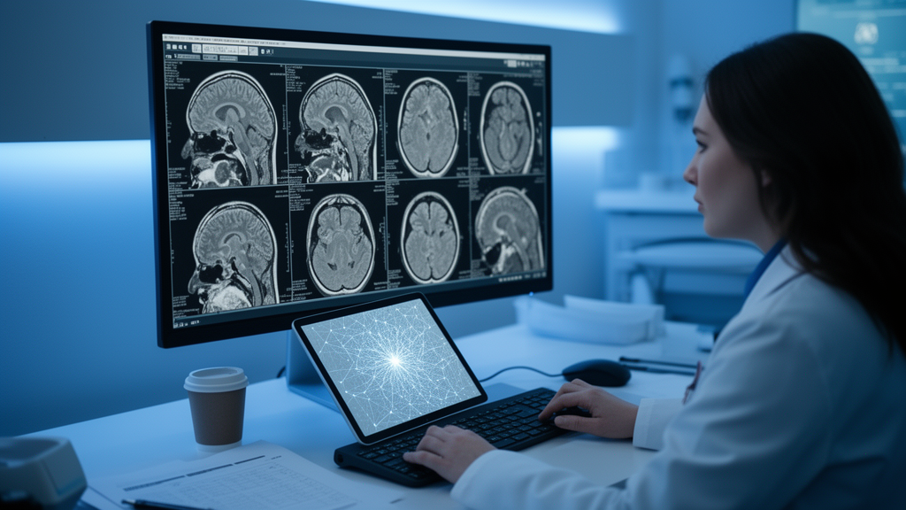
The team, led by Edouarde Assumpta Tsafack and Ghislain Feudjio, describes the framework in a study published in the journal Discover Imaging on 10 September 2026. Rather than relying on the massive deep learning networks that dominate modern medical image analysis, the approach extracts a small set of handcrafted mathematical features from a tiny patch of each scan and feeds them into a shallow neural network, keeping the computational burden low enough for hospitals that lack expensive hardware.
The motivation is practical. Stroke is a leading cause of death and disability worldwide, responsible for roughly 6.6 million fatalities each year, and about half of all survivors live with chronic disability. Rapid treatment depends on distinguishing ischemic stroke, caused by blocked blood flow to the brain and accounting for about 87 percent of cases, from hemorrhagic stroke, marked by sudden bleeding in the brain and making up roughly 13 percent. Computed tomography is often the first imaging tool used because it is cost-effective, widely accessible and good at revealing intracranial lesions, but interpreting scans quickly and accurately remains a challenge, particularly in resource-constrained settings where the study’s authors work.
The pipeline begins with standardization. The team converted raw DICOM files to Hounsfield Units using scanner metadata, resampled the images to a uniform 1.0 by 1.0 millimeter resolution following the Image Biomarker Standardization Initiative, and applied a re-segmentation range of minus 10 to 90 Hounsfield Units to isolate soft tissue and stroke lesions. Intensities were discretized into fixed bins for texture analysis. An expert radiologist then manually outlined the boundaries of pathological tissue, and the region of interest was standardized as a circular patch with an 8-pixel radius, equivalent to 8 millimeters on the resampled images, producing a 17 by 17 pixel matrix of 289 pixels centered on the abnormality.
From each of these small patches the method extracts exactly eight numbers, split evenly between two complementary domains. Four features come from the discrete wavelet transform, using the Daubechies 4 mother wavelet at a single level of decomposition: the mean of the low-frequency approximation sub-band, which captures the overall structure and most of the image’s energy, plus the energy of the horizontal, vertical and diagonal detail sub-bands. The other four come from the gray-level co-occurrence matrix, a second-order statistical texture descriptor computed over eight gray-level bins, averaged across four angular directions at a one-pixel displacement: energy, contrast, correlation and homogeneity. Together these quantify both coarse intensity patterns and fine spatial texture, the difference between the dense, bright appearance of pooled blood in a hemorrhage and the subtle darker changes of early infarction.
Those eight values, scaled between zero and one, feed a small multilayer perceptron with an 8-neuron input layer, a hidden layer of 32 neurons, a second hidden layer of 16 neurons and a 3-neuron softmax output giving probabilities for hemorrhagic, ischemic and normal tissue. Training used the Adam optimizer with a starting learning rate of 0.001, categorical cross-entropy loss, dropout regularization and early stopping. The clinical data came from 66 patients at the Regional Hospital Center of Bafoussam in sub-Saharan Africa, scanned on a Siemens SOMATOM Emotion 16 scanner, with 22 patients in each of the three classes and five to six continuous CT slices per subject, yielding 348 slices in total.
A crucial design choice guards against inflated results: the evaluation used a strict patient-grouped five-fold cross-validation, in which all slices from any single patient stayed within one partition, so the model was always tested on entirely new anatomy. Under this protocol the combined wavelet-and-texture model reached a mean accuracy of 99.17 percent, with a 95 percent confidence interval of 97.54 to 99.72 percent. The confusion matrix was nearly diagonal, with all 116 hemorrhagic cases correctly classified and only three misclassifications total between the ischemic and normal classes. Calibration curves for all three classes lay almost on the line of perfect calibration, with Brier scores between 0.000 and 0.005, meaning the model’s stated confidence closely matched its actual hit rate.
The ablation results show why the hybrid design matters. A baseline model without engineered features managed only 32.49 percent mean accuracy, barely above the 33 percent chance level for a three-class problem. Texture features alone lifted accuracy to 57.09 percent, but ischemic and normal scans remained heavily confused, with 33 ischemic cases predicted as normal and 49 normal cases predicted as ischemic. A statistical analysis explains the gap: Kruskal-Wallis tests across all 348 slices found that the wavelet-derived mean and energy features had effect sizes near 0.89, meaning class membership explained about 89 percent of their variance, while the texture features reached only about 0.33. In other words, the frequency-domain measurements carry most of the discriminative signal, particularly for separating ischemic tissue from healthy parenchyma.
The framework also held up on external data. On the public Teknofest-2021 dataset, which contains hemorrhagic, ischemic and normal CT slices, the same preprocessing and region extraction produced a stable benchmark accuracy of 98.00 percent under stratified image-level cross-validation, with strong F1-scores across the five folds. Benchmarking against a simple k-nearest-neighbor classifier on the same feature set, which achieved 98.30 percent on the clinical data, suggested that the dual-domain representation itself, rather than any particular classifier, drives the performance. In literature comparisons the approach matched or exceeded several deep learning models, including SqueezeNet, MobileNet and OZNet variants, and came close to a MaxViT architecture on the Teknofest data, though the authors note that a ViT and GAN-based method reported a higher peak accuracy of 99.06 percent.
The team also built a prototype graphical interface that runs on standard CPU hardware. It is deliberately not a fully automated detector: a clinician or radiologist manually points to the suspicious region on a CT slice, and the system then extracts the wavelet and texture features in real time and returns an immediate classification with a confidence score. Tested on unseen images from the Teknofest external-test folder, the tool highlighted localized lesions with attached confidence levels. The authors position this as a second-reader system that can draw radiologist attention to subtle abnormalities during high-volume screening, supporting rather than replacing human expertise, and they emphasize that the full feature-level design makes the model’s decisions transparent in a way that deep networks often are not.
The researchers are candid about the limits. The system depends on manual localization of the region of interest by an expert, its performance hinges on the isotropic resampling step, and the clinical cohort comes from a single hospital with a modest number of patients, so broader validation is needed before any clinical use. Planned next steps include automated suggestions of candidate lesion regions, multi-center validation across different scanners, testing alternative wavelet families, inter-observer repeatability trials, and extending the feature extraction from 2D slices to 3D lesion volumes. For now, the study stands as a proof of concept that a carefully engineered eight-number fingerprint, rather than a heavyweight neural network, can carry most of the diagnostic weight in stroke CT classification, an appealing proposition for emergency departments where computing power, electricity and specialist time are all in short supply.
Subject of Research: A lightweight machine learning method for classifying stroke types in brain CT scans
Article Title: A lightweight hybrid feature extraction method for stroke segmentation, classification, and prediction in brain CT scans using multilayer perceptron
Article References: Tsafack, E. A., Jiazet, E., Pone, J. R. M., Djimeli-Tsajio, A. B., & Feudjio, G. (2026). A lightweight hybrid feature extraction method for stroke segmentation, classification, and prediction in brain CT scans using multilayer perceptron. Discover Imaging, 3(1), Article 9. https://doi.org/10.1007/s44352-026-00030-9
Image Credits: AI Generated
DOI: 10.1007/s44352-026-00030-9
Keywords: stroke, brain CT, machine learning, discrete wavelet transform, gray-level co-occurrence matrix, multilayer perceptron, computer-aided diagnosis, medical imaging, radiomics, ischemic stroke, hemorrhagic stroke, resource-constrained settings
News Source: Cassandra Pierce. (October 28, 2026). Lightweight AI Tool Classifies Stroke Types from Brain CT Scans. Scienmag.



