


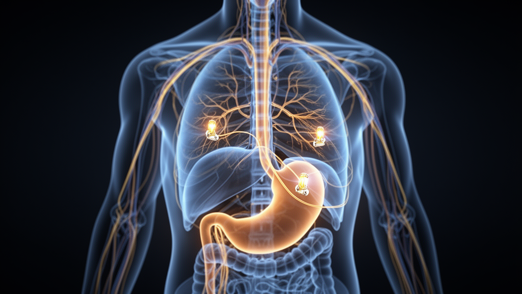
When the NIH Common Fund launched the SPARC program in 2015, few bioelectronic medicine approaches were routinely used in clinical care. Researchers lacked detailed knowledge of how peripheral nerve pathways innervate internal organs, had a limited understanding of the neural signaling patterns that control organ function, and had no neural interfaces optimized for the small-diameter autonomic nerves that measure less than 500 micrometers across. A decade later, the program has officially ended, and a new review published in Bioelectronic Medicine takes stock of what it delivered: more than $350 million awarded to nearly 100 projects, over 500 publications, and more than 300 datasets, all aimed at catalyzing therapies that modulate electrical activity in the autonomic nervous system to improve organ function.
The review, led by Bhavesh Patel and colleagues including Maryann E. Martone, organizes the program’s legacy around four interdependent focus areas: anatomical and functional mapping, technology development, translational research, and data and modeling infrastructure. The autonomic nervous system, comprising the parasympathetic and sympathetic divisions, controls every internal organ besides the brain, which is why the authors argue that the clinical reach of bioelectronic medicine could extend across neurological, cardiovascular, urological, gastrointestinal, respiratory, metabolic, and other conditions. Chronic diseases are a leading cause of morbidity, with 43 percent of people in North America experiencing two or more chronic conditions, a burden that motivates the search for therapies beyond drugs and irreversible surgery.
On the mapping front, Phase 1 of the program concentrated on five major organ systems: the heart, lungs, stomach, colon, and bladder. Funded teams used tools such as optogenetics and genetic labeling to characterize neural circuitry under healthy conditions, then moved to large animal and disease models to inform translational work. Additional projects mapped the innervation of less-studied organs including the spleen, pancreas, esophagus, kidney, liver, adipose tissue, bone, and bone marrow, and in partnership with the NIH HEAL Initiative, some teams studied peripheral circuits involved in visceral pain signaling.
Among the mapping initiative’s notable outcomes was a comprehensive three-dimensional atlas of the rat intrinsic cardiac nervous system at single-cell resolution, built by researchers at the University of Central Florida and Thomas Jefferson University. The atlas characterized how intrinsic cardiac neurons are distributed between individuals and across sexes, and the team developed computational approaches that bridge molecular-level gene expression data with sparse cellular-level electrophysiology to probe the functional role of this nerve network in cardiac regulation. Meanwhile, investigators at the University of Wisconsin-Madison and Duke University combined microdissection, histology, and immunohistochemistry to compare the structure of the cervical and subdiaphragmatic vagus nerve in humans, swine, and rats. They found significant species differences, including greater morphological variability across human samples than in swine or rats, and went on to build models simulating vagus nerve stimulation in all three species. Those models showed that applying the same stimulation parameters across individuals or across species produced a large range of nerve responses, underscoring the need for systematic approaches that account for individual- and species-specific differences.
Phase 2 shifted the mapping focus squarely onto the human vagus nerve, the longest nerve in the body and the autonomic conduit for parasympathetic control over most internal organs. In the REVA project, investigators at the Feinstein Institute for Medical Research, Case Western Reserve University, and Duke University used gross dissection and multi-scale imaging of cadaveric vagus nerves to generate high-resolution connectivity profiles, quantifying nerve diameter, fascicle count, fascicle area, and perineurium thickness, and describing the extent of anatomical variability among individuals. The parallel VESPA clinical study is capturing multi-organ readouts of standardized stimulation parameters in patients implanted with vagus nerve stimulation devices, seeking to determine how best to stimulate specific nerve fibers for specific therapeutic effects. Some of these datasets are already public on the SPARC Portal.
The technology initiative produced tools that have, in several cases, already reached the clinic or the market. A team at Cleveland Clinic, Case Western Reserve University, and the Louis Stokes Cleveland VA Medical Center developed the Urological Monitor of Conscious Activity, a sensor implanted into the bladder lumen of conscious ambulating animals that measures pressure and volume for up to 30 days. Using it, the investigators showed that high-frequency sacral nerve stimulation can reduce pelvic floor and urethral contraction, enabling bladder emptying. The bladder technology has been licensed to Bright Uro, Inc., which has received FDA approval for its initial device, the Glean, for improved diagnosis of lower urinary tract dysfunction in people. Separately, researchers at the California Medical Innovations Institute developed Fecobionics, a wireless, FDA-cleared simulated feces device that measures pressures, orientation, bending, and shape changes during defecation, offering an objective assessment of anorectal disorders including constipation and fecal incontinence.
Computational modeling formed a third pillar. Duke University researchers built ASCENT, an open-source pipeline for anatomically realistic modeling of electrical stimulation of peripheral nerves, which has since been updated with Python-based fiber simulation, neural recording capability, and redesigned documentation. Multiple studies have used it to analyze and design neuromodulation therapies, from validated human, pig, and rat vagus nerve stimulation models to inter-species translation of stimulation parameters and comparisons of cuff versus intraneural electrodes. At the IT’IS Foundation, researchers leveraged the Sim4Life platform with dosimetric assessment, induced heating simulation, and electrochemical damage predictors to perform in silico safety assessment of sufficient rigor to serve as regulatory evidence; multiple major manufacturers of bioelectronics implants are now using this pipeline for submissions to the FDA.
Phase 2 also turned toward open-source, clinical-grade hardware. Through the SPARC HORNET program, the CARSS team built the OpenNerve device, a modular implantable pulse generator with four leads supporting up to eight channels of current-controlled nerve stimulation and up to eight channels of biosensing, including electrical, mechanical, and enzyme-free electrochemical sensors for acetylcholine, catecholamines, pH, and temperature. The COSMIIC team developed the Networked Neuroprosthesis with leads for muscle stimulation and circuits for high-frequency nerve block. Both teams’ libraries include regulatory documentation, hardware designs, manufacturing processes, and firmware designed to comply with FDA requirements, intended to lower the financial and regulatory barriers to new clinical-grade devices.
On the translational side, Phase 1 funded pre-clinical and clinical proof-of-concept studies. Indiana University researchers showed in a canine model that high-intensity subcutaneous nerve stimulation was more beneficial than vagus nerve stimulation in treating atrial fibrillation, though low-intensity stimulation showed a proarrhythmic effect requiring further investigation. A University of Michigan team demonstrated in a feline model that closed-loop sacral nerve stimulation, which reads bladder sensory signals from the dorsal root ganglia and modulates stimulation in real time, outperformed open-loop approaches, boosting bladder capacity and cutting required stimulation time by more than half. At Johns Hopkins, spinal cord stimulation at the thoracic level normalized gastric motility in a canine model of gastroparesis, and a Purdue University-led collaboration validated a method of measuring vagal compound action potentials through the skin of patients implanted with gastric electrical stimulators, finding that specific response signatures predict improvements in specific gastroparesis-related symptoms. In Phase 2, a Neuromodulation Prize Competition rewarded selective neuromodulation approaches capable of independently regulating two or more autonomic functions, with first place going to a University of Pittsburgh multichannel implantable device for sacral-pudendal neuromodulation addressing bladder, bowel, and sexual disorders, and second place to Juniper Biomedical’s micro-implantable system for stress urinary incontinence, overactive bladder, and fecal incontinence.
Underpinning everything is the SPARC Data and Resource Center, whose four cores at the University of Pennsylvania, the University of California San Diego, the IT’IS Foundation, and the University of Auckland built the SPARC Portal around a standardized data structure endorsed by the International Neuroinformatics Coordinating Facility. As of February 2026, the portal hosts 414 datasets and over 86 terabytes, including 308 research datasets, 55 computational models, and 51 anatomical models, with more than 115,000 dataset views and 15,000 downloads between February 2023 and December 2025. The o2S2PARC cloud modeling platform now counts over 2,700 registered users and has found adoption beyond neuromodulation, in brain stimulation planning, exposure safety assessment, and medical imaging. The authors are candid about remaining barriers to translation: the need for accurate and reliable sensors, long-term stability of implanted devices, and robust algorithms that can accommodate changing neural outputs, along with the far less controlled conditions of real-world use. Still, with the program officially ended in 2025 and activities continuing through no-cost extensions and data sharing, the SPARC Portal remains open to submissions from other funded projects, and the infrastructure is already supporting NIH HEAL, Epilepsy.Science, and European projects, a pathway the authors argue will keep catalyzing new neuromodulation therapies long after the program itself has closed.
Subject of Research: Outcomes of the NIH SPARC bioelectronic medicine program
Article Title: Accelerating the development of bioelectronic medicine: outcomes and innovations from the NIH SPARC initiative
Article References: Patel, B., Pikov, V., Damaser, M. S., Boline, J., Tappan, S. J., Nickerson, D. P., Ward, M. P., Gunalan, P. K., Neufeld, E., Ludwig, K. A., Grethe, J. S., Wagenaar, J. B., & Martone, M. E. (2026). Accelerating the development of bioelectronic medicine: outcomes and innovations from the NIH SPARC initiative. Bioelectronic Medicine, 12(1), Article 20. https://doi.org/10.1186/s42234-026-00213-z
Image Credits: AI Generated
DOI: 10.1186/s42234-026-00213-z
Keywords: bioelectronic medicine, NIH SPARC, autonomic nervous system, neuromodulation, vagus nerve, open science, SPARC Portal, neural interfaces, data sharing, computational modeling, peripheral nerve stimulation, Accelerating
News Source: Denise Maddox. (October 28, 2026). NIH’s SPARC Program Ends After a Decade of Mapping Nerves and Building Open Bioelectronic Tools. Scienmag.



