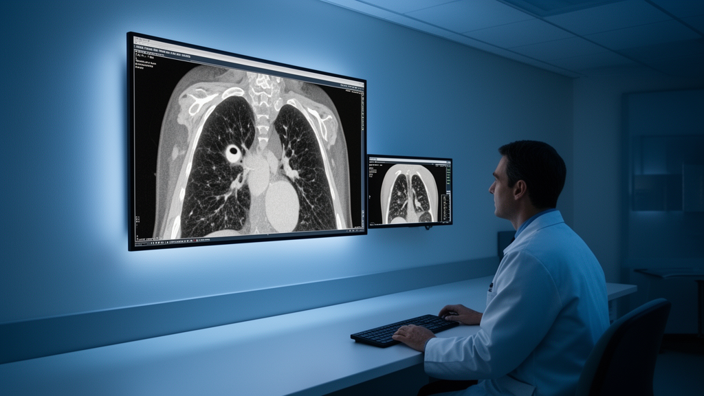
A sweeping map of the early clinical evidence on photon-counting detector CT suggests the technology can render pulmonary nodules more clearly than conventional detectors while exposing patients to measurably less radiation, but the review’s authors stop well short of calling for routine adoption. The scoping review, published in European Radiology Cardiovascular and Thoracic, identified just five studies, covering 318 patients and 20 cadaveric human lungs, that have evaluated pulmonary nodule visualization on the only commercially available whole-body photon-counting system. Within that small and heterogeneous body of work, a consistent picture emerges: better subjective image quality, more morphologic detail visible in subsolid nodules, and dose reductions of roughly 12 to 18 percent in same-day comparisons against energy-integrating detector CT.
Pulmonary nodules are among the most common findings on chest CT, appearing in incidental scans, lung cancer screening programs, and oncologic surveillance. Detection alone is rarely the deciding factor in patient management. Under the Lung-RADS and Fleischner Society frameworks, radiologists and referring physicians weigh features such as spiculation, lobulation, pleural retraction, internal attenuation, air bronchograms, and margin definition to estimate malignancy risk and choose between surveillance, biopsy, or resection. How sharply a scanner renders those features therefore has direct clinical consequences, particularly for subsolid nodules, where the size and attenuation of any solid component drive the recommendation.
Conventional energy-integrating detector CT has well-characterized limitations that bear on exactly this task. Scintillator-based detectors convert X-rays into visible light before digitization, embedding electronic noise in the output signal, and contrast-to-noise ratio degrades as radiation dose falls. Energy averaging weights high-energy photons despite their lower soft-tissue contrast, blunting sensitivity to ground-glass attenuation differences. Reflective septa separating detector elements impose a practical floor on pixel size, limiting the depiction of submillimeter features. Photon-counting detectors, which entered clinical practice in 2021, address each constraint differently: semiconductor sensors made of cadmium telluride or cadmium-zinc-telluride convert individual photons directly into electrical pulses whose amplitude encodes photon energy, an adjustable energy threshold rejects electronic noise, and the absence of septa permits ultra-high-resolution acquisition.
The review team, led by Felix Barajas Ordonez and Felipe Aluja-Jaramillo, followed JBI scoping review methodology and PRISMA-ScR reporting standards, searching MEDLINE/PubMed, Embase, and Scopus from January 2021 through January 2026. Two reviewers screened records independently, achieving 90 percent agreement in a calibration exercise and a kappa of 0.82 for full-text screening. Consistent with scoping review methodology, no formal risk-of-bias assessment was performed. From 160 initial records, the search ultimately yielded five studies published between 2023 and 2025, conducted in the USA, China, Japan, and Switzerland, all using the same NAEOTOM Alpha platform from Siemens Healthineers.
The dose findings are among the review’s most concrete results. In one same-day comparison at screening-equivalent doses, photon-counting CT delivered a volume CT dose index of 0.61 versus 0.73 mGy on conventional CT, roughly 16 percent lower, with significantly better diagnostic quality and sharpness scores. At diagnostic-range doses, a second same-day study reported CTDIvol of 4.28 versus 4.86 mGy, about 12 percent lower, and effective dose of 1.79 versus 2.17 mSv, about 18 percent lower, again with superior subjective image quality. A retrospective comparison found photon-counting CTDIvol of 2.97 versus 6.90 mGy on prior conventional scans, though that comparison involved different scanners and protocols. A fourth study, comparing two dose levels within the photon-counting scanner itself, cut the dose by roughly 83 percent, from 0.65 to 0.11 mGy, a level comparable to a chest radiograph.
That ultra-low-dose tier produced the review’s most striking detection result. In 172 nodules of 5 mm or larger, semiautomated detection maintained an overall sensitivity of 0.94 at the chest-radiograph-equivalent dose, with subgroup sensitivity of 0.95 to 0.96 for solid nodules and 0.85 to 0.90 for subsolid nodules; the nodules that were missed were significantly smaller. Lung-RADS classification between the two dose levels showed Cohen’s kappa of 0.62 to 0.76 and accuracy of 0.78 to 0.85. The review’s authors argue that because photon-counting detectors’ principal physical advantage is electronic-noise suppression, that advantage is expressed most fully at screening-equivalent doses, where electronic noise dominates the noise budget, and compresses at diagnostic doses where quantum noise prevails.
Morphologic characterization showed some of the largest reported gaps between the two detector technologies. In the same-day diagnostic-dose study of exclusively subsolid nodules, photon-counting CT detected significantly higher rates of air cavities, vessel signs, spiculation, and pleural retraction, and identified additional morphologic features in 34 to 73 percent of subsolid nodules compared with conventional CT performed the same day. A retrospective study of 203 pathologically classified subsolid nodules found higher detection of heterogeneous attenuation, lobulation, bubble-like sign, air bronchogram, pleural indentation, and vascular sign with ultra-high-resolution photon-counting acquisition. At screening-equivalent doses, nodule border visualization scores favored photon-counting CT, with the largest effect for ground-glass nodules.
The cadaveric work adds both support and a cautionary note. In 20 inflated human lungs with histological correlation, photon-counting CT at a 1024-pixel matrix achieved the highest morphological concordance with histology, and full concordance scores were reached only at that matrix size, with a median minimum detectable nodule diameter of 604 micrometers. Yet when the same specimen set was re-analyzed against ultra-high-resolution conventional CT, the minimum detectable diameter was 604 micrometers for conventional CT at 2048 matrix versus 680 micrometers for photon-counting CT, a difference that was not statistically significant, and the conventional reconstruction actually outperformed photon-counting CT for airways larger than 1000 micrometers. The authors read this as evidence that advanced reconstruction on conventional detectors can match or exceed detector-driven gains for selected endpoints, meaning observed advantages reflect detector physics, reconstruction algorithms, and acquisition protocol jointly rather than detector technology alone.
The review is candid about its limits. All five studies were single-center evaluations on a single commercial platform, no study was prospectively registered, and no investigation tested whether the visualization gains translate into improved diagnostic accuracy or patient outcomes. Nodule populations differed substantially across cohorts, with two studies enrolling subsolid nodules exclusively and one evaluating predominantly solid nodules, and no study stratified results by age, sex, or body mass index. Because the two cadaveric reports analyzed the same specimen set, they cannot be pooled with in vivo data, and cadaveric conditions lack cardiorespiratory motion, patient habitus, and clinical workflow. The authors also note that since Lung-RADS categories were derived from conventional CT data, those categories should not yet be upgraded solely on morphologic features seen only on photon-counting images.
What the evidence does support, the authors conclude, is a measured case for prioritizing photon-counting CT, where it is available, for low-dose screening protocols and subsolid-nodule characterization, the settings where electronic-noise suppression matters most. The priority next steps they identify are multicenter prospective same-day trials at screening-equivalent doses against ultra-high-resolution conventional CT with deep-learning reconstruction, head-to-head benchmarking of photon-counting CT combined with artificial intelligence against AI on conventional CT, multi-vendor data, and clinical-outcome endpoints. Until such studies are done, the review frames photon-counting CT as a promising but unproven tool for nodule evaluation, one whose early results are encouraging precisely because the field has not yet subjected them to the comparative-effectiveness testing that implementation would demand.
Subject of Research: Image quality and morphologic visualization of pulmonary nodules with photon-counting detector CT
Article Title: Pulmonary nodule image quality and morphologic visualization with photon-counting detector CT: a scoping review
Article References: Barajas Ordonez, F., Safar, I. E. M., Rendón-Garavito, A. M., Yüksel, C., Kuhl, C., Pantoja-Burbano, O. A., & Aluja-Jaramillo, F. (2026). Pulmonary nodule image quality and morphologic visualization with photon-counting detector CT: a scoping review. European Radiology Cardiovascular and Thoracic, 1(1), Article 4. https://doi.org/10.1007/s44449-026-00004-3
Image Credits: AI Generated
DOI: 10.1007/s44449-026-00004-3
Keywords: photon-counting CT, pulmonary nodules, image quality, radiation dose, subsolid nodules, Lung-RADS, lung cancer screening, CT detectors, scoping review, cadaveric lungs, morphologic characterization, ultra-low dose
News Source: Ophelia Keating. (October 28, 2026). Photon-counting CT sharpens lung nodule images at lower radiation doses, review finds. Scienmag.