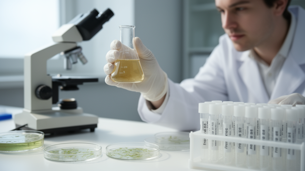
A team of chemists and marine biologists in Italy has developed a simplified, seedless method for making silver nanoparticles and shown in laboratory cultures that the particles strongly suppress the growth of two widespread marine diatoms, Cylindrotheca closterium and Skeletonema marinoi. The work, published in the journal Discover Metals, was motivated by the persistent problem of biofouling: the unwanted accumulation of microalgae and other organisms on boat hulls, ships and other submerged human-made structures, which raises hydrodynamic resistance, increases fuel consumption and can help spread invasive species.
The synthesis itself is deliberately minimalist. The researchers mixed a solution of polyvinylpyrrolidone, a common polymer stabiliser known as PVP, with silver nitrate under continuous stirring, then added dilute ascorbic acid, ordinary vitamin C, drop by drop to trigger the reduction of silver ions to metallic silver. The solution turned from colourless to an intense brown as the nanoparticles formed. What makes the protocol distinctive is that ascorbic acid served as the sole reducing agent. In most colloidal syntheses, ascorbic acid is used alongside stronger reducing agents to fine-tune reduction kinetics; here the team relied entirely on its moderate reduction potential to drive both nucleation and growth, avoiding the aggressive and toxic reagents that similar procedures typically require.
The word seedless, the authors caution, does not mean the reaction escapes nucleation-driven growth, which is intrinsic to any liquid-phase synthesis. It simply means that no preformed exogenous seeds were added to the reaction mixture. Their one-pot protocol involves nucleation in situ followed by growth, distinguishing it from the seed-mediated methods that dominate laboratory nanoparticle production. According to the paper, the complete synthesis took only about ten minutes, with the ascorbic acid added dropwise until the characteristic yellow-brown colour signalled nanoparticle formation. The entire process was carried out in autoclaved water and finished with a purification combining centrifugation and filtration, a step the researchers describe as crucial not only for removing metal precursors, residual reductant and excess polymer but also for eliminating bacterial contaminants that could compromise the biological assays that followed.
Characterisation of the resulting material, dubbed AgNP@PVP, rested on three complementary techniques. Ultraviolet-visible spectroscopy showed a strong, well-defined absorption band at 425 nanometres, the surface plasmon resonance that the authors call unequivocal spectroscopic evidence of colloidal silver nanoparticle formation. The position and symmetric, single shape of the band are consistent with spherical metallic silver particles in the nanometre range, although the team notes the data cannot exclude the coexistence of silver oxide or surface-oxidised silver species. Transmission electron microscopy confirmed the isotropic shape: well-dispersed spheres with a diameter of about 20 plus or minus 4 nanometres. Infrared spectroscopy verified the PVP coating. The carbonyl stretching band of pure PVP at 1653 wavenumbers shifted to 1643 in the presence of the nanoparticles, and new bands appeared near 1436, 1091 and 1041 wavenumbers, features the authors attribute to perturbations of the polymer backbone and to donor-acceptor or charge-transfer interactions between the electron-rich oxygen and nitrogen atoms of the pyrrolidone ring and the silver surface. These interactions, they argue, are the molecular basis of the steric stabilisation that keeps the particles from clumping.
The stability of the suspension was checked in both freshwater and saltwater, the medium in which the particles would later be dispersed with the microalgae, over a period of 96 hours. The concentration of PVP adsorbed on the particle surfaces was estimated from the same purified sample by measuring the polymer’s characteristic carbonyl absorbance around 200 nanometres against a calibration curve, yielding an estimate of 1.5 times ten to the minus two molar.
For the biological tests, the team selected two diatoms representing different ecological strategies and microalgal families. Cylindrotheca closterium is a pennate, benthic species known for its ability to form biofilms, while Skeletonema marinoi is a centric, planktonic species. Both strains originally came from the Adriatic Sea and were maintained at the Stazione Zoologica Anton Dohrn culture collection. Cultures were grown in Guillard’s F/2 medium prepared with sterile, filtered seawater at 37 parts per thousand salinity, and each treatment began with an inoculum of 5000 cells per millilitre.
The experimental design separated the effect of the silver core from that of the coating polymer. Seven conditions were tested for each species: an untreated control, three concentrations of pure PVP solution alone, and three concentrations of the AgNP@PVP suspension. The nanoparticle stock was extremely dilute, 1.7 times ten to the minus ten molar, and the exposure conditions were chosen to reflect environmentally realistic scenarios for nanoparticle release from antifouling coatings in marine benthic environments. Cultures were kept in triplicate at 19 degrees Celsius under a 12-hour light and 12-hour dark cycle at 100 micromoles of photons per square metre per second, and cell density was counted daily for eight days in a Sedgwick-Rafter chamber under a light microscope after fixation with Lugol’s solution.
The results were unambiguous. Both species in the control cultures were still in stationary phase at day seven, while the nanoparticle-treated algae showed severe reductions in cell viability. At the highest nanoparticle concentration, algae entered the decline phase by day three and were already dead by day seven; at the medium concentration they entered decline at day six. The numbers tell the story. Cylindrotheca closterium reached a final concentration of only 5 times ten to the third cells per millilitre at the highest dose, 8 times ten to the third at the medium dose and 2 times ten to the fifth at the lowest dose, against roughly 6 times ten to the fifth in the control. Skeletonema marinoi fared similarly, peaking at 5 times ten to the third, 6 times ten to the fourth and 7 times ten to the fourth cells per millilitre at the high, medium and low doses respectively, compared with about 2 times ten to the fifth in controls. The differences were reported as statistically significant, with p values below 0.001 from student’s t-tests across three biological replicates. Crucially, cultures exposed to PVP alone showed no adverse effects at any concentration, maintaining cell densities comparable to the untreated control, which the authors say confirms that the inhibition is an intrinsic property of the silver component rather than the coating. PVP, they note, is a passive coating essential for preventing aggregation but biologically inert in this assay.
Supporting visual evidence reinforced the counts. In representative flasks, control cultures of C. closterium showed a dense sedimentation of algae at the bottom, while the nanoparticle-treated vials remained completely clear with no detectable cell deposits, which the authors describe as direct evidence of growth inhibition. Light microscopy also revealed that treated C. closterium cells exposed to the medium nanoparticle dose were shorter than controls, a change consistent with earlier reports that stress conditions can alter this species’ growth and cell size. Skeletonema marinoi chains looked similar to controls in shape, although more free-living bacteria were observed associated with them at the medium dose. At the highest nanoparticle concentration both algae died, while the lowest dose induced no particular changes in cell shape.
The authors are careful to frame the scope of what was and was not demonstrated. A direct correlation between particle size, plasmon resonance peak and antifouling activity was not examined and would require dedicated experiments, which they plan alongside tests of nanoparticles with different surface coatings and sizes and assessments of toxicological effects on other marine organisms. That caution matters because silver nanoparticles are known from the literature to exert ecotoxicological effects on non-target aquatic organisms, including bacteria, crustaceans and fish, and because traditional antifouling coatings based on copper and organotin compounds such as TBT already carry a heavy legacy of environmental persistence, non-specific toxicity, bioaccumulation and resistant fouling species. Any real-world application, the team writes, must balance efficacy against fouling algae with environmental safety, possibly through coatings designed for controlled release of the particles. They call for acute and chronic toxicity tests and evaluations of bioaccumulation and persistence in the marine environment before any field use. Within those limits, the study delivers a rapid, inexpensive, seedless route to stabilised silver nanoparticles and controlled-laboratory evidence that, at very low concentrations, they can arrest the proliferation of two ecologically distinct marine diatoms, a promising first step toward nanoparticle-based alternatives to conventional antifouling chemistry.
Subject of Research: Green synthesis of PVP-stabilized silver nanoparticles and their inhibition of marine diatom growth
Article Title: Green synthesized silver nanoparticles act as algal growth inhibitors
Article References: Candreva, A., Shoukat, T., Lauritano, C., Ruffolo, S. A., Russa, M. F. L., Santillán-Sarmiento, A., & Deda, M. L. (2026). Green synthesized silver nanoparticles act as algal growth inhibitors. Discover Metals, 59(1), Article 6. https://doi.org/10.1007/s13404-026-00375-3
Image Credits: AI Generated
DOI: 10.1007/s13404-026-00375-3
Keywords: silver nanoparticles, green synthesis, ascorbic acid, PVP, marine diatoms, Cylindrotheca closterium, Skeletonema marinoi, antifouling, biofouling, algae growth inhibition, nanotechnology, Discover Metals
News Source: Denise Maddox. (October 28, 2026). Seedless silver nanoparticles curb growth of two marine diatoms in lab tests. Scienmag.