


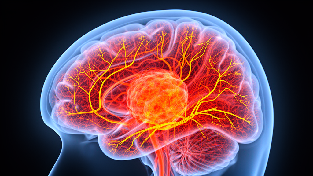
Arterial spin labelling, an MRI method that uses magnetically labelled blood as its own contrast agent, has shown a strong correlation with microvessel density in brain tumours, according to a prospective study from Mansoura University in Egypt. In a cohort of 37 adults with newly diagnosed primary brain tumours, the researchers found that relative tumour blood flow measured by three-dimensional arterial spin labelling correlated positively with microvessel density assessed by histopathology, with a correlation coefficient of 0.742 and a p-value of 0.001. Mean tumour blood flow, calculated directly within the lesion without normalisation, also correlated with microvessel density, but more weakly, with a coefficient of 0.378. Both correlations remained statistically significant after the researchers applied a Bonferroni correction for multiple comparisons, and the relative tumour blood flow was the only variable that retained statistical significance in a multivariate regression analysis that also accounted for patient age, tumour type and tumour location.
The study, published in the Egyptian Journal of Radiology and Nuclear Medicine, recruited patients from the neuro-oncology outpatient clinic at Mansoura University Hospitals between December 2024 and February 2026. All participants were over 18 years old and newly diagnosed with primary brain tumours. The investigators excluded patients with recurrent tumours, metastatic disease, or previous chemotherapy or radiotherapy. The cohort comprised 14 males and 23 females, aged 18 to 66 years, and included 16 intra-axial tumours, 15 extra-axial tumours and 6 posterior fossa and skull base tumours. Microvessel density was measured after surgical excision or stereotactic biopsy using CD34 immunohistochemistry, a marker of endothelial progenitor cells that play a major role in angiogenesis.
Arterial spin labelling differs fundamentally from conventional contrast-enhanced MRI. While gadolinium-based post-contrast imaging captures the static leakage of contrast across a disrupted blood-brain barrier, perfusion imaging tracks the dynamic passage of blood through the brain’s microvasculature. The technique uses magnetically labelled blood as an endogenous tracer, requiring subtraction of labelled and control images with a delay between tagging and image readout. Because it requires no exogenous contrast agent, arterial spin labelling is suitable for patients with renal impairment or hypersensitivity to contrast media, and previous studies have reported diagnostic accuracy comparable to dynamic susceptibility contrast methods for assessing primary and metastatic brain tumour perfusion.
For the imaging protocol, the team used a 1.5 Tesla Siemens MAGNETOM Aera scanner with pseudocontinuous arterial spin labelling, chosen for its high signal-to-noise ratio and superior labelling efficiency compared with other variants. The sequence employed a post-labelling delay of 1020 milliseconds, four background suppression pulses suppressing about 95 percent of static tissue signal, and an acquisition time of three minutes. Motion artefacts were corrected using the SAMER technique, which relies on a rapid low-resolution scout scan lasting three to five seconds. The researchers placed a standardised region of interest of roughly 50 square millimetres over the most densely perfused part of each tumour, avoiding haemorrhage, cystic degeneration and necrosis, and averaged three such regions. Relative tumour blood flow was then calculated by dividing this average by the mean of three regions placed over the most perfused part of the contralateral cortical grey matter, or the cerebellar hemisphere for infratentorial and skull base lesions.
Reproducibility of the measurements was tested by having the same reader, blinded to histopathology, repeat the analysis after a minimum of four weeks. Intra-class correlation coefficients were 0.995 for mean tumour blood flow and 0.983 for relative tumour blood flow, and Bland-Altman analysis showed excellent intra-observer agreement. The histopathological analysis was performed by an experienced pathologist blinded to the imaging diagnosis, following the hotspot method originally described by Weidner: three areas with the highest vessel count were selected at low power, and microvessels were counted at high power, with the average taken as the microvessel density value expressed in vessels per high-power field.
The correlation between relative tumour blood flow and microvessel density held across the pooled sample regardless of tumour location, which was the study’s primary aim. A post hoc power analysis using a bivariate normal model, a correlation coefficient of 0.742, an alpha error of 0.05 and the sample of 37 patients indicated a statistical power of 99.8 percent for the primary correlation. The authors note, however, that this post hoc analysis does not compensate for the limited sample size in subgroup analyses. The multivariate regression, which included relative tumour blood flow, patient age, tumour type and location as variables, confirmed that relative tumour blood flow was the only significant predictor of microvessel density, with a p-value of 0.001.
The study also explored, as an exploratory aim, whether perfusion parameters correlated with tumour grade within individual tumour groups. In the intra-axial group, there was a borderline positive correlation between relative tumour blood flow and grade, with a p-value of 0.046 and a coefficient of 0.504, but this did not survive Bonferroni correction, which set the corrected threshold at 0.016. No statistically significant correlation was found between relative tumour blood flow and grade in the extra-axial group, where the p-value was 0.872, or in the posterior fossa and skull base group, where it was 0.407. Mean tumour blood flow showed no significant correlation with grade in any of the three groups. The authors caution that the study was not powered for these subgroup analyses and that the absence of a significant correlation does not indicate a true lack of biological association, particularly given the small size of individual groups, which ranged from 6 to 16 patients.
The findings carry several important caveats acknowledged by the authors. The use of CD34 alone, which cannot distinguish original tumour vessels from neovessels stimulated by angiogenesis, may have led to overestimation of microvessel density, and the reported values likely reflect total vascularity rather than angiogenic activity alone. The hotspot approach, selecting both the point of highest perfusion on imaging and the areas of highest vessel count on histology, may have introduced systematic bias that artificially inflated the correlation coefficient. The workstation lacked a three-dimensional region-of-interest feature, so two-dimensional regions susceptible to partial volume effects were used despite the volumetric acquisition, and absolute cerebral blood flow values in standard units could not be calculated, meaning the mean tumour blood flow values were reported in arbitrary units. The study was also single-centre, included only adults, did not perform interobserver reliability testing, and lacked access to the molecular biomarkers of the 2021 WHO classification, such as 1p/19q co-deletion and H3 K27M, limiting the subcategorisation of certain tumours.
Despite these limitations, the authors argue that incorporating arterial spin labelling into multiparametric MRI for pre-operative brain tumour assessment offers practical benefits. Relative tumour blood flow provides insight into the microperfusion of gliomas, which has been described as a predictor of tumour grade, and perfusion maps may help differentiate true brain neoplasms from mimics such as tumefactive demyelination, potentially reducing the need for invasive stereotactic biopsies. In meningiomas, identifying highly perfused lesions could help flag tumours that might benefit from pre-operative embolization. The authors conclude that arterial spin labelling perfusion parameters, particularly relative tumour blood flow, exhibit a strong correlation with microvessel density and angiogenesis in brain tumours, but they stress that the technique should be used cautiously in pre-operative assessment, combined with other advanced neuroimaging techniques, and validated in larger, more homogeneous future cohorts before its role in tumour grading can be established.
Subject of Research: Correlation between arterial spin labelling MRI perfusion parameters and microvessel density in brain tumours
Article Title: Correlation between three-dimensional arterial spin labelling perfusion parameters and microvessel density in brain tumours: a prospective study
Article References: Ayyad, M. S., El-Toukhy, N. A. E.-G., Elkashef, W., Shahein, M., & Mansour, M. (2026). Correlation between three-dimensional arterial spin labelling perfusion parameters and microvessel density in brain tumours: a prospective study. Egyptian Journal of Radiology and Nuclear Medicine, 57(1), Article 232. https://doi.org/10.1186/s43055-026-01900-2
Image Credits: AI Generated
DOI: 10.1186/s43055-026-01900-2
Keywords: arterial spin labelling, brain tumours, microvessel density, MRI perfusion, angiogenesis, relative tumour blood flow, CD34 immunohistochemistry, gliomas, meningiomas, posterior fossa tumours, pre-operative imaging, non-invasive biomarker
News Source: Nathaniel Bowman. (October 28, 2026). MRI Blood-Flow Technique Tracks Tumour Vessel Density Without Contrast Agents. Scienmag.



