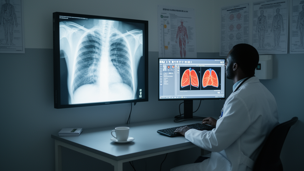
Researchers in Ethiopia have built a deep learning system that classifies chest X-rays into three categories — normal, pneumonia, and tuberculosis — using images collected from public hospitals in the Amhara region. After systematic optimization, their customized VGG19 model reached 99% accuracy, precision, recall, and F1-score on internal test data, with an area under the receiver operating characteristic curve (AUC) of 0.9993. When tested on an independent external dataset of 3,568 images, performance dropped to 88.83% accuracy and a macro F1-score of 83.92%, a decline the authors attribute to domain shift between datasets rather than model failure.
The study, published in Discover Artificial Intelligence, addresses a pressing clinical gap. Pneumonia remains a leading cause of illness and death worldwide, and tuberculosis continues to affect millions, with an estimated 10.6 million new cases reported in 2021 according to World Health Organization figures cited by the researchers. In Ethiopia, and in the Amhara region in particular, these respiratory diseases impose a heavy healthcare burden, worsened by limited access to skilled radiologists. Manual interpretation of chest X-rays can take a day or longer, delaying diagnosis and treatment, and traditional diagnostic pathways combining clinical evaluation and microbiological testing are time-consuming and prone to human error.
To build the dataset, the team gathered 2,100 chest X-ray images from five healthcare facilities, including Debre Markos Comprehensive and Specialized Hospital, Gondar University Hospital, Dessie Referral Hospital, Bahir Dar Felege Hiwot Hospital, and Sanitas Medium Clinic. Ground-truth labels were assigned by radiologists during routine clinical practice, and images were sorted into Normal, Pneumonia, and Tuberculosis folders based on radiological assessment and clinical records. The classes were balanced, with equal numbers of images in each category, which the researchers note resolved the data imbalance problem that often undermines medical image classifiers. Because the dataset was retrospective and anonymized, information on the number of radiologists involved and inter-rater agreement statistics was unavailable, so the study relied on the original clinical labels.
Preprocessing played a substantial role in the pipeline. Images were resized to a uniform 224 by 224 pixels and pixel values were rescaled to the range zero to one. Training data underwent augmentation with rotations up to 10 degrees, width and height shifts of 0.1, zooming by a factor of 0.1, brightness adjustment between 0.8 and 1.2, and random contrast changes, with a nearest-fill mode covering empty regions created by the transformations. Validation and test images were only rescaled, preserving their original distribution for unbiased evaluation. The authors report that comparing performance with and without augmentation showed clear improvements across accuracy, precision, recall, and F1-score, along with more stable training and reduced fluctuation in validation loss.
Six architectures were evaluated under identical conditions: a baseline Sequential convolutional neural network trained from scratch, plus five pre-trained transfer learning models — VGG19, MobileNetV3-Large, EfficientNetV2-B0, EfficientNetV2-B1, and InceptionResNetV2. At baseline, VGG19 led with 82% accuracy and macro-averaged precision, recall, and F1-score of 81%, while the Sequential CNN reached 79.23% accuracy and InceptionResNet 70%. MobileNetV3-Large and the EfficientNetV2 variants performed considerably lower, with accuracies between 48% and 60%, which the authors suggest may reflect underfitting or insufficient adaptation to the dataset.
Optimization then transformed the results. The team applied a two-stage transfer learning strategy: in the first stage, the pre-trained convolutional backbone was frozen and only the newly added classification layers were trained for ten epochs using the Adam optimizer with a learning rate of 0.001, early stopping, and plateau-based learning rate reduction. In the second stage, the top convolutional layers were unfrozen and training continued at a much lower learning rate of 0.00001 to refine high-level features without disrupting earlier learned representations. Hyperparameters were tuned empirically — dropout rates from 0.3 to 0.7 were tested with 0.4 performing best, dense layer sizes from 128 to 1024 neurons were evaluated with 256 proving optimal, and learning rates of 0.001, 0.0001, and 0.00001 were compared. Class weighting addressed residual imbalance, and callbacks including EarlyStopping, ReduceLROnPlateau, and ModelCheckpoint supported stable convergence.
The customized VGG19 that emerged uses Global Average Pooling, a smaller first fully connected layer, and dropout, reducing parameters relative to the original architecture while preserving its deep stack of small 3-by-3 convolutional filters. Early convolutional layers were frozen to avoid catastrophic forgetting of general visual features, while deeper layers were fine-tuned to the medical domain. After optimization, VGG19 achieved 0.990 plus or minus 0.001 across accuracy, precision, recall, and F1-score, and an AUC of 0.9993 plus or minus 0.0003, with narrow confidence intervals indicating stable, reproducible performance. The Sequential CNN followed at 98% across metrics, MobileNetV3-Large and EfficientNetV2-B1 reached 96 to 97%, and InceptionResNet and EfficientNetV2-B0 landed at 95 to 97% with AUC values of 0.996 to 0.997. The authors attribute the improvement from 82% to 99% to the refined training strategy rather than any change in data or evaluation, since both experiments used identical datasets, splits, preprocessing, and metrics. Statistical analysis using bootstrap confidence intervals from 1,000 resamples and pairwise McNemar tests confirmed that the customized VGG19 significantly outperformed baseline architectures, and repeated training with multiple random seeds yielded consistent results.
External validation provided a more sobering picture. The final model was tested on an entirely separate Kaggle dataset of 3,568 chest X-rays containing 719 Normal, 1,500 Pneumonia, and 1,349 Tuberculosis cases. Accuracy fell to 88.83%, precision to 86.35%, recall to 88.83%, F1-score to 83.92%, and AUC to 0.8865 — a drop of 10.17 percentage points in accuracy. Class-wise, Normal cases performed best with an F1-score of 0.9231 and sensitivity of 93.46%, followed by Pneumonia at 0.8726 and Tuberculosis at 0.8437, with tuberculosis showing the highest misclassification rate, likely because its radiographic patterns overlap with pneumonia. The researchers argue this degradation is consistent with published reports in medical AI, where external validation typically reveals more realistic performance estimates than internal testing, and they emphasize that the internal results likely represent optimistic estimates. Differences in acquisition protocols, patient populations, image quality, and equipment may all have contributed.
Several limitations deserve attention. Because patient-level identifiers were unavailable, data were split at the image level, creating a potential risk of data leakage if multiple images from the same patient appeared in different subsets — a risk the authors mitigated through the external validation, bootstrap intervals, and repeated seeding, but which future studies should address with patient-level partitioning. Hospital identifiers were also absent, preventing scanner-specific subgroup analysis. Notably, the VGG19 model received extensive optimization while the MobileNet and EfficientNet variants were trained with standard settings, so performance comparisons may reflect unequal tuning effort. All experiments ran on CPU-only hardware — an Intel Core i3 processor with 4 GB of RAM and no dedicated GPU — which ruled out k-fold cross-validation and illustrates the resource constraints the work is meant to address. The customized VGG19 contained roughly 20 million parameters at about 77 MB, while the lighter models ranged from 14 to 59 MB and offered faster CPU inference, highlighting a trade-off between representational power and computational efficiency for resource-constrained environments.
Explainability remains an open question. The team used Gradient-weighted Class Activation Mapping, or Grad-CAM, with anatomically constrained lung-field masks to visualize which image regions drove predictions, observing attention concentrated in peripheral lung regions for Normal cases, lower lung regions for Pneumonia, and upper lung regions for Tuberculosis. However, no independent radiologist review of these heatmaps was performed, and the authors explicitly caution that the visualizations represent model attention patterns rather than clinically verified evidence of disease localization. They acknowledge that post-hoc explainability methods can produce plausible-looking but misleading attention maps, and they state that structured radiologist review with quantified agreement measures will be required before any claims about clinical relevance can be made. Future work will also pursue prospective multicenter validation, patient-level evaluation, larger multi-hospital datasets, lateral chest X-rays, and additional pulmonary diseases. Alongside the research, the team built a Streamlit web application prototype that lets users upload chest X-ray images and receive instant feedback, positioned as a decision-support tool rather than a deployed diagnostic system. The findings suggest that carefully optimized deep learning models, even trained on modest hardware, could support automated screening for pneumonia and tuberculosis in settings where radiologists are scarce — provided that independent validation remains the yardstick for real-world performance.
Subject of Research: Deep learning classification of pneumonia and tuberculosis from chest X-rays in Ethiopia
Article Title: Pneumonia and tuberculosis detection and classification using deep learning in resource limited settings, Ethiopia
Article References: Flatie, A. B., Wordofa, Z. R., Mengistu, A. K., Gedefaw, A. E., Bitew, K. G., Abebe, A. W., Yitayew, A. M., & Emiru, E. D. (2026). Pneumonia and tuberculosis detection and classification using deep learning in resource limited settings, Ethiopia. Discover Artificial Intelligence, 6(1), Article 1003. https://doi.org/10.1007/s44163-026-02068-4
Image Credits: AI Generated
DOI: 10.1007/s44163-026-02068-4
Keywords: deep learning, pneumonia, tuberculosis, chest X-ray, VGG19, transfer learning, Ethiopia, medical imaging, convolutional neural networks, Grad-CAM, external validation, resource-limited settings
News Source: Blake Davidson. (October 28, 2026). Deep learning model reads chest X-rays for pneumonia and tuberculosis in Ethiopia. Scienmag.