Sepsis after major burn injury affects between 20.1 and 39.1 percent of patients and stands as the leading cause of death among those who survive the initial injury, according to a narrative review published in October 2026 by specialists in burns critical care. The review, which synthesised literature identified through a comprehensive search of PubMed, Medline, Embase, Scopus and Web of Science spanning 1998 to April 2026, argues that the syndrome presents clinicians with a uniquely tangled problem: the very physiological storm triggered by thermal injury mimics the pathological changes of sepsis itself, blurring the line between expected post-burn response and life-threatening infection.
The scale of the underlying problem is considerable. An estimated 13 million severe burns occurred worldwide in 2021, and the World Health Organisation estimates around 180,000 people die each year as a consequence of burn injury. That burden is projected to rise with population growth and ageing, and will principally affect low- and middle-income countries. Against this backdrop, the review describes why burn-related sepsis resists the diagnostic frameworks that serve other critically ill populations, and why management depends less on any single test than on a holistic, multidisciplinary reading of each patient’s trajectory.
The pathophysiology explains much of the diagnostic confusion. Severe burn injury disrupts the skin barrier extensively, but it also triggers release of catecholamines, cortisol and pro-inflammatory cytokines including tumour necrosis factor-alpha, interleukin-1 and interleukin-6. These mediators promote endothelial dysfunction, glycocalyx shedding and increased vascular permeability, contributing to organ dysfunction even in the absence of infection. The hyperinflammatory, hypermetabolic response raises energy expenditure, cardiac output, body temperature and oxygen consumption. Overlapping this is a dynamic period of immunosuppression marked by impaired T-cell function, altered cytokine signalling, reduced antigen presentation, altered neutrophil activity and reduced antimicrobial peptide production, compounded by microbiome disruption and polymicrobial colonisation during often prolonged intensive care admissions.
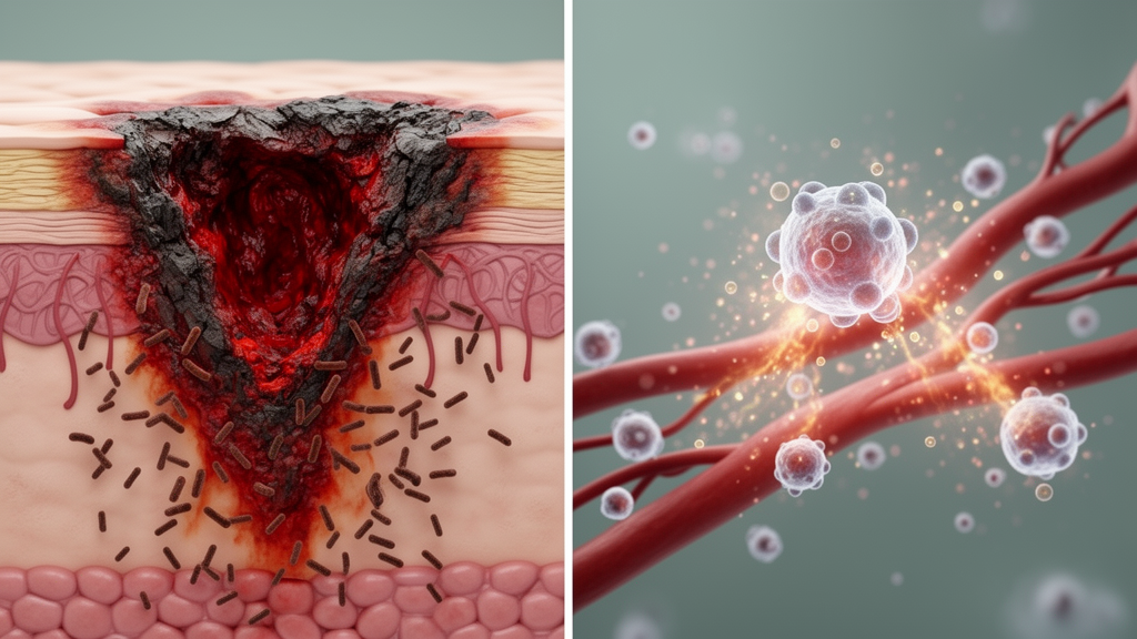
Burn wounds are the most common source of infection, with a continuum running from superficial colonisation to invasive infection and systemic burn wound sepsis. Invasive infection occurs when microorganisms penetrate viable tissue surrounding the burn eschar, allowing bloodstream entry and systemic dissemination. Skin graft donor sites can also serve as infection sources. Beyond wounds, prolonged intensive care stays and invasive devices heighten susceptibility to ventilator-associated pneumonia, catheter-related bloodstream infections, catheter-associated urinary tract infections and intra-abdominal infections. Distinguishing infection from colonisation is a central challenge, because burn wounds are routinely colonised and positive wound cultures alone do not confirm invasive disease.
Standard definitions fare poorly here. Many burn patients fulfil systemic inflammatory response syndrome criteria—fever, tachycardia, tachypnoea and leukocytosis—in the absence of infection, and a systemic inflammatory response is almost universal in burns exceeding 30 percent of total body surface area. The Sepsis-3 definition, which identifies sepsis through a rise of two or more points in the Sequential Organ Failure Assessment score, has improved recognition in general intensive care populations, but the physiological response to severe burn injury frequently influences components of that score, significantly reducing its specificity. The American Burn Association has proposed burn-specific criteria requiring clinical suspicion of infection plus at least three of six predictors, including extreme temperature changes, progressive tachycardia, tachypnoea, thrombocytopenia, hyperglycaemia and intolerance of enteral feeding. Notably, no single scoring system has been demonstrated to have adequate accuracy to serve as a diagnostic standard.
Biomarkers tell a similarly sobering story. A recent systematic review and meta-analysis incorporating 28 studies, 57 biomarkers and 1,517 subjects found white cell count to be the least helpful conventional biomarker, with poor sensitivity of 47 percent and moderate specificity of 65 percent. C-reactive protein, which correlates with burn size, shows high sensitivity of 86 percent but poor specificity of 54 percent as a single measurement; however, a prospective study of 60 adults with major burns found that a rising serum CRP predicted sepsis with 93 percent sensitivity and 80 percent specificity. Procalcitonin offered an area under the receiver operating characteristic curve of 0.79, with 73 percent sensitivity and 75 percent specificity, and a rise of more than 2 nanograms per millilitre from baseline has been recommended as a trigger for considering sepsis. Kinetics, in short, outperform absolute thresholds.
Emerging markers remain research tools for now. Pancreatic stone protein performed similarly to procalcitonin in a prospective study of 90 adults with burns covering at least 15 percent of total body surface area, reaching a maximum area under the curve of 0.89 at day seven. Complete blood count derivatives showed only modest predictive value in a cohort of 2,757 burn patients, with areas under the curve just above 0.65. Neutrophil extracellular trap markers showed promising discrimination in a 63-patient observational study. For fungal infection, circulating Mucorales DNA screening twice weekly in patients with burns over 15 percent of total body surface area was associated with reduced mortality in a single-centre study, later confirmed in a bicentric study of patients with burns of at least 30 percent, with a median lead time of 11 days before positive cultures. Yet no biomarker has been validated in a randomised trial specific to burn patients.
The microbial landscape shifts over the patient’s hospital course. Early infections, in the first three days, are dominated by Gram-positive organisms from the patient’s own skin and mucosal microbiota, particularly Staphylococcus aureus and beta-haemolytic streptococci, though isolates at this stage often represent commensal colonisers rather than true pathogens. Gram-negative bacteria from the gastrointestinal tract and intensive care environment emerge during days four to seven, and after the first week nosocomial, often multidrug-resistant pathogens prevail, including Pseudomonas aeruginosa, Acinetobacter baumannii and Enterobacterales such as Klebsiella pneumoniae. Fungal infections and viral reactivation typically emerge later, with Candida species the most common fungal pathogens and non-albicans species increasingly reported. Resistance mechanisms including extended-spectrum beta-lactamases and carbapenemase-producing Enterobacterales are increasingly reported, driven by the intense antimicrobial pressure characteristic of burn units.
Management rests on established pillars rather than novel agents. Routine prophylactic systemic antibiotics are not recommended, as they promote multidrug-resistant organisms without improving outcomes; infection prevention instead relies on bundled measures including hand hygiene, contact precautions, single-occupancy rooms with negative pressure ventilation for intensive care patients, and meticulous water quality control. Early tangential excision and grafting—the removal of necrotic eschar, an ideal bacterial culture medium, typically within seven days—has been the single most important advance in reducing mortality from burn wound sepsis. A 2021 meta-analysis of six randomised trials including 512 patients suggested this strategy may reduce mortality and hospital stay while increasing transfusion requirements, though the certainty of these treatment effects was considered low. Antibiotic stewardship demands attention to altered pharmacokinetics, as increased volume of distribution and enhanced renal clearance can produce sub-therapeutic levels requiring therapeutic drug monitoring.
The review also tempers enthusiasm for experimental approaches. Corticosteroid use in major burns was independently associated with significantly increased odds of mortality, multi-organ failure and sepsis in a small retrospective study of 22 patients, though this should not preclude low-dose hydrocortisone for septic shock; oxandrolone’s apparent mortality benefit in one single-centre retrospective study has not been replicated, and the drug lost Food and Drug Administration approval in 2023. Immunomodulatory therapies such as recombinant interleukin-7 and checkpoint inhibitors have shown promise mainly in pre-clinical models, polymyxin B hemoperfusion lacks robust evidence of survival benefit in burns, and bacteriophage therapy has so far demonstrated reduced bacterial counts only in mice. The authors conclude that effective identification and management of burn sepsis will continue to rely on assiduous infection prevention, prompt surgical excision and judicious antibiotic stewardship, with emerging diagnostics and therapies likely years away from routine practice.
Subject of Research: Diagnosis and management of sepsis following major burn injury
Article Title: Sepsis in the major burn patient
Article References: McGovern, C., Paton, L., Martin-Loeches, I., Dumont, M., Dépret, F., Stoppe, C., Depetris, N., Casaer, M., & Puxty, K. (2026). Sepsis in the major burn patient. Critical Care: Sepsis and Severe Infection, 1(1), Article 16. https://doi.org/10.1186/s44541-026-00019-1
Image Credits: AI Generated
DOI: 10.1186/s44541-026-00019-1
Keywords: sepsis, burns, critical care, biomarkers, antimicrobial resistance, infection control, Sepsis-3, procalcitonin, burn wound infection, multidrug-resistant organisms, surgical excision, immunomodulation
News Source: Ophelia Keating. (October 28, 2026). Why Sepsis in Major Burn Patients Defies Standard Diagnosis and Treatment. Scienmag.