A 75-year-old man walked into a respiratory clinic with lungs clouded by ground-glass opacities and consolidation spread across both fields on computed tomography, yet he reported no symptoms at all. He had never smoked, reported no exposure to dust or other environmental hazards, and his medication history was unremarkable. His oxygen saturation measured 89 percent on room air, a level low enough to signal that something serious was unfolding inside his chest even before he felt it. Laboratory results offered few clues: C-reactive protein was a near-normal 0.09 mg/dL, Krebs von den Lungen-6, a marker of lung injury, stood at 225 U/mL, and serum proteinase 3 anti-neutrophil cytoplasmic antibody was 12.8 U/mL. There were no physical findings pointing to an autoimmune disease, no symptoms of heart failure, and N-terminal pro-brain natriuretic peptide was a reassuring 45 pg/mL. Even tumor markers seemed quiet, with carcinoembryonic antigen at 2.0 ng/mL and soluble interleukin-2 receptor at 171 U/mL.
What followed was a diagnostic race against a rapidly deteriorating patient, and a case that its physicians now report as a cautionary tale about how pancreatic cancer can wear the mask of interstitial lung disease. The clinical team performed bronchoalveolar lavage promptly, retrieving fluid that was slightly turbid and pale yellow, with a return of just 41 percent. Analysis found neither pathogens nor malignant cells. The total cell count was 3.0 × 10^5 cells/mL, composed of 87 percent macrophages, 11 percent lymphocytes, and 2 percent neutrophils, with a CD4/CD8 ratio of 2.17. But the patient’s oxygenation worsened during the examination, so a biopsy could not be performed. With his respiratory condition and radiologic findings deteriorating rapidly after the procedure, the team suspected rapidly progressive interstitial lung disease of unknown cause.
Treatment began with methylprednisolone at 1000 mg/day for three days as pulse therapy. It failed. Oxygenation and the radiological opacities did not improve, so two additional courses of methylprednisolone were given, followed by a reduction to oral prednisolone at 60 mg/day. At the worst point, the patient required 100 percent concentrated oxygen delivered through a high-flow nasal cannula. Intravenous cyclophosphamide at 500 mg/m² was then added, administered every two weeks for a total of three doses starting three weeks after the initial treatment. Prednisolone was subsequently tapered by 5 to 10 mg every three to five days. Then something remarkable happened: follow-up CT showed a reduction in the ground-glass opacities and consolidation in both lungs. Nine weeks after the initial treatment, the patient was discharged without oxygen therapy, taking 20 mg/day of prednisolone.
The improvement, however, was an illusion. Several days after discharge, the man returned to the emergency department with dyspnea. CT revealed new consolidation and ground-glass opacities in both lungs layered on top of the pre-existing lesions, along with a left pleural effusion, but nothing highly suggestive of malignancy, no lymphadenopathy and no mass lesions. Magnetic resonance imaging told a different story elsewhere in the body, revealing dilatation and disruption of the main pancreatic duct. Blood tests followed suit: duodenal pancreatic cancer antigen 2 was 870 U/mL, SPan-1 antigen was 15 U/mL, and carbohydrate antigen 19-9 was below 2 U/mL. Sputum cytology identified adenocarcinoma, and lung metastases from pancreatic adenocarcinoma were diagnosed on the basis of the radiological findings and cytology.
By then the patient was too debilitated for active cancer treatment and received only palliative care. He died, and an autopsy was performed. The autopsy thus explained the entire clinical course: two overlapping processes, one malignant and one inflammatory, occupying the same chest and responding in overlapping ways to the same drugs.
The central puzzle of the case is why the lungs improved at all under treatment aimed at interstitial lung disease. Cyclophosphamide, the drug added when steroids alone failed, possesses both anti-tumor and immunosuppressant effects. Although it is not commonly used in current standard chemotherapy regimens for advanced pancreatic cancer, a study investigating its role as maintenance therapy for pancreatic adenocarcinoma found it may be effective as a standalone agent. In this patient, the temporary improvement in pulmonary opacities may have resulted from prednisolone and/or cyclophosphamide acting on the organizing pneumonia and possibly on the lung metastases themselves, although the physicians note that distinguishing between these two entities was challenging. The same drug was, in effect, treating two entirely distinct diseases at once.
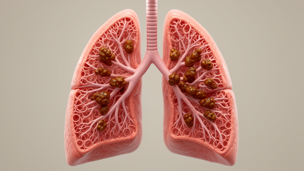
The biology behind the masquerade is well documented in the literature. Lung metastases of pancreatic cancer are reported in 15 to 50 percent of cases and are not uncommon. On CT, they take many forms, including various patterns of nodules, ground-glass opacities, and consolidation. Alveolar patterns, in particular ground-glass opacities and consolidation, suggest a lepidic growth pattern in lung metastases of pancreatic cancer, exactly what the autopsy later confirmed in this patient. Typically, these metastases present as isolated, asymmetrical, or multifocal lesions. Only rarely do they appear as bilateral diffuse lung opacities that mimic interstitial lung disease, which is precisely what made this case so difficult to unmask before irreversible time and treatment had been spent.
The relationship between interstitial lung disease and cancer runs in both directions, and the case report situates itself within this broader picture. It is well known that cancer can develop from fibrotic interstitial lung disease, but cancer can also cause various patterns of interstitial lung disease. In this case, however, autopsy pathology did not reveal carcinomatous lymphangiopathy. Instead, it identified pancreatic ductal carcinoma and organizing pneumonia. Organizing pneumonia is a known complication of treatments including chemotherapy and immunotherapy, and it may also occur in association with the cancer itself. It has been reported to complicate not only lung cancer but other malignant tumors such as leukemia and gastrointestinal cancers.
One further caveat deserves emphasis. Several studies have suggested that secondary organizing pneumonia, including tumor-related organizing pneumonia, and cryptogenic organizing pneumonia may have similar treatment responses. But the evidence remains limited by the small number of reported cases of tumor-related organizing pneumonia, so definitive conclusions cannot be drawn. This matters for clinicians weighing whether a response to immunosuppression rules out an underlying malignancy. In this patient, the response to cyclophosphamide and prednisolone was real but temporary, and it reflected partial activity against both an inflammatory process and an untreated cancer rather than confirmation of an interstitial lung disease diagnosis.
The lesson the authors draw is direct and practical. Even when diffuse bilateral ground-glass opacities and consolidation respond to treatment for interstitial lung disease, lung metastasis from extrapulmonary malignancies remains a critical differential diagnosis and should be carefully considered when planning therapeutic strategies for undiagnosed lung disease. The case also illustrates a structural dilemma: when a patient is deteriorating fast enough that treatment must start before a diagnosis is secured, drugs with pleiotropic effects such as cyclophosphamide can blur the very signal clinicians rely on to judge whether their working diagnosis was correct. A temporary improvement in bilateral diffuse opacities, in other words, is not proof of interstitial lung disease. It may be the quiet success of a drug working against a cancer no one has yet named, in a patient whose window for finding out is closing.
Subject of Research: A case report of pancreatic adenocarcinoma lung metastases mimicking interstitial lung disease and temporarily responding to cyclophosphamide and corticosteroid treatment.
Article Title: Lung Metastases of Pancreatic Adenocarcinoma Masquerading as Temporary Improvement of Bilateral Diffuse Lung Opacities Following Treatment for Interstitial Lung Disease
Article References: Kusunoki, S., Tasato, M., Sokai, A., Iwata, T., Hayashi, Y., Yasuda, N., Kashiwagi, I., & Nishimura, T. (2026). Lung Metastases of Pancreatic Adenocarcinoma Masquerading as Temporary Improvement of Bilateral Diffuse Lung Opacities Following Treatment for Interstitial Lung Disease. Respirology Case Reports, 14(10), Article e70795. https://doi.org/10.1002/rcr2.70795
Image Credits: AI Generated
DOI: 10.1002/rcr2.70795
Keywords: pancreatic adenocarcinoma, lung metastases, interstitial lung disease, cyclophosphamide, organizing pneumonia, ground-glass opacities, case report, misdiagnosis, lepidic growth, autopsy, palliative care, bronchoalveolar lavage
News Source: Nathaniel Bowman. (October 27, 2026). Pancreatic Cancer Hiding in the Lungs: A Case of Mistaken Interstitial Disease. Scienmag.