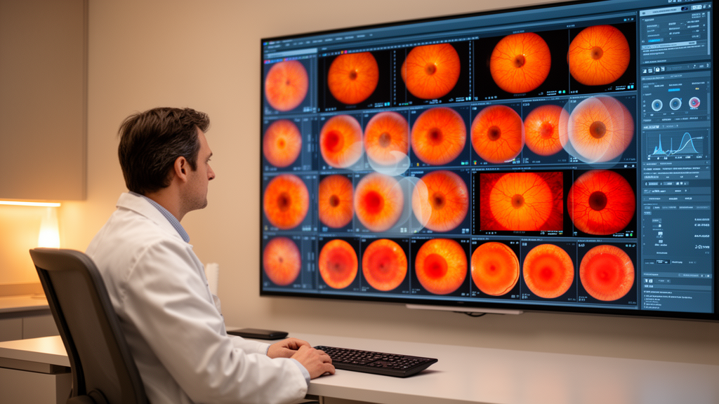
A research team from Putian First Hospital in China now reports a single deep learning framework, called MyoSpectrumNet, designed to carry out all of these jobs at once, using retinal photographs drawn from several very different data sources. Most existing artificial intelligence studies in this field have concentrated on one narrow task at a time, trained on one dataset at a time. The publicly available fundus image collections relevant to pathologic myopia offer complementary information, but their labels only partially overlap, which has made it difficult to build one model that serves all purposes.
MyoSpectrumNet was built specifically to bridge that gap. The framework was developed using three established datasets: HPMI, PALM and MMAC. Together these sources cover the spectrum of pathologic myopia assessment, from a simple yes-or-no screening decision, through grading of myopic maculopathy on established severity scales, to an auxiliary estimate of a patient's spherical equivalent refraction and the delineation of specific myopia-related lesions in the retina.
Rather than training a separate network for each job, the researchers created a source-conditioned multi-task checkpoint, meaning the model is told which data source an image comes from so it can adapt to the different labelling conventions of each collection. Performance was evaluated on held-out partitions from the known sources, with the model never tested on images it had seen during training. For the primary comparisons, the team used single-view inference, meaning each image was assessed once in its standard form.
A more elaborate four-view test-time augmentation, in which predictions are averaged across multiple transformed versions of an image, was retained only as a sensitivity analysis within the MyoSpectrumNet framework itself, so that headline results would not be inflated by extra inference machinery unavailable to the comparison models. The screening results were strikingly strong. Under single-view inference, task-specific baseline models achieved an area under the curve of 0.987 for detecting pathologic myopia in the HPMI dataset, while MyoSpectrumNet reached 0.985 on the same test.
On the PALM dataset the baselines scored 0.996 and the unified model 0.993. For grading myopic maculopathy severity, measured by quadratic weighted kappa, both approaches scored identically at 0.907. In other words, folding four tasks into one network cost almost nothing in screening and grading accuracy.
The auxiliary tasks told a more nuanced story. A dedicated ConvNeXt-T squeeze-and-excitation baseline estimated spherical equivalent refraction with a mean absolute error of 1.006 dioptres, with a 95 percent confidence interval of 0.873 to 1.147 dioptres, and a Pearson correlation of 0.801 with true values. MyoSpectrumNet's single-view estimate was less precise, with a mean absolute error of 1.259 dioptres.
Conversely, on lesion segmentation the unified framework came out ahead: a DeepLabV3-ResNet50 baseline achieved an overall Dice coefficient of 0.397, with a 95 percent confidence interval of 0.324 to 0.468, while the MyoSpectrumNet interpretation checkpoint reached 0.472 under single-view inference. The authors are careful about how these cross-model comparisons should be read. The auxiliary outputs of the original MyoSpectrumNet training runs were preserved only as aggregate summaries, which means the refraction and lesion comparisons are descriptive rather than definitive head-to-head trials.
The team explicitly states that no universal claim of task-specific superiority is being made. What the results do demonstrate is the central trade-off of joint learning: combining tasks in one model did not improve every output, yet it preserved a set of clinically complementary capabilities that would otherwise require several separate systems. Important limitations remain.
The complete multi-output framework has not yet been externally validated on a dataset entirely unseen during development, a step that is essential before any diagnostic AI can be trusted in real-world clinics. All reported figures come from held-out partitions of the same three source collections, and performance on new cameras, new populations or different stages of disease remains an open question. The study, published in Frontiers in Medicine in the ophthalmology section, positions MyoSpectrumNet as a unifying foundation for pathologic myopia assessment rather than a finished clinical product.
Subject of Research: A multi-task deep learning framework for assessing pathologic myopia from heterogeneous fundus image datasets
Article Title: MyoSpectrumNet: a unified deep learning framework for pathologic myopia spectrum assessment using heterogeneous multi-source fundus datasets
Article References: Zhang, B., Lin, F., Li, W., & Lin, B. (2026). MyoSpectrumNet: a unified deep learning framework for pathologic myopia spectrum assessment using heterogeneous multi-source fundus datasets. Frontiers in Medicine, 13, 1881289. https://doi.org/10.3389/fmed.2026.1881289
Image Credits: AI Generated
DOI: 10.3389/fmed.2026.1881289
Keywords: pathologic myopia, deep learning, fundus photography, myopic maculopathy, multi-task learning, screening, lesion segmentation, spherical equivalent, HPMI, PALM, MMAC, ophthalmology
News Source: Ophelia Keating. (October 27, 2026). One AI Model Tackles the Full Spectrum of Pathologic Myopia From Fundus Photos. Scienmag.