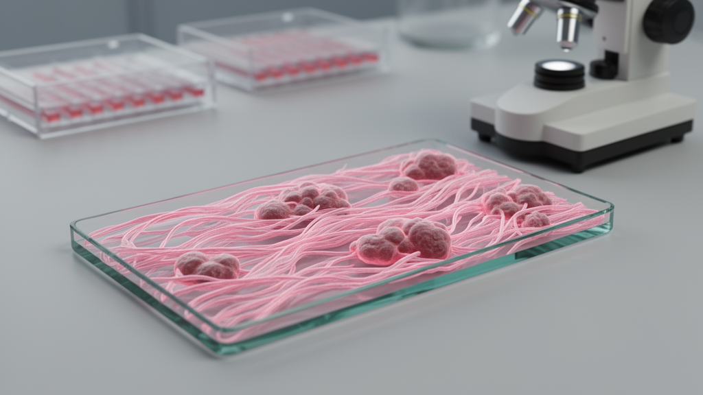
Collagen architecture within the tumour microenvironment has emerged as an important structural biomarker in cancer, but measuring it reliably has typically required Second Harmonic Generation (SHG) microscopy, a label-free technique that visualizes fibrillar collagen in detail. Despite its power, SHG imaging remains constrained by high cost, specialized instrumentation and a limited field of view, which have kept it out of routine clinical practice. A new proof-of-concept study asks whether the collagen features that SHG captures can instead be approximated from ordinary, digitally scanned histopathology slides.
The researchers worked with formalin-fixed, paraffin-embedded breast tumour tissues, including both benign samples and invasive ductal carcinoma (IDC) specimens with varying collagen content. Each matched region of interest was analysed in two ways: by SHG microscopy, and by whole-slide brightfield imaging of sections stained with Masson–Goldner’s Trichrome, a routine laboratory stain that highlights collagen. The stained slides were then quantified using two independent digital image analysis approaches, a conventional ImageJ-based workflow called TWOMBLI and a machine learning-based computational pipeline.
Before any comparison, the team confirmed that SHG signals could be consistently detected from the Trichrome-stained sections, establishing that the two modalities were imaging compatible material. They then quantified collagen structural parameters, including collagen deposition area, fibre number and alignment metrics, and compared the values across imaging modalities using correlation analysis.
The quantitative comparison demonstrated significant concordance between SHG-derived collagen metrics and those obtained from the digital analysis pipelines, with the strongest agreement seen for collagen area and fibre alignment. In other words, computationally extracted features from routine stained slides tracked closely with the label-free optical measurements of the same collagen architecture.
The authors present these results explicitly as a proof of concept: computational analysis of routine histopathological images can capture key spatial features of collagen organization comparable to SHG microscopy. Because whole-slide imaging and digital pathology are already widely deployed, the approach could in principle be scaled without the specialized instrumentation that SHG demands, making extracellular matrix assessment more accessible in clinical settings.
The study stops short of clinical deployment. It was conducted on a defined set of breast tissue samples, and the concordance was strongest for specific parameters rather than every collagen metric. Even so, the findings suggest a potentially scalable and clinically accessible route to quantifying extracellular matrix architecture in tumour tissues, positioning collagen organization as a biomarker that could be assessed from slides already generated in standard diagnostic workflows.
Subject of Research: Quantitative collagen architecture assessment from routine histopathological images compared with Second Harmonic Generation microscopy in breast tumour tissues
Article Title: Quantitative assessment of collagen architecture from routine histopathological images shows concordance with Second Harmonic Generation microscopy
Article References: Ingawale, V., Dandapat, K., Konkada Manattayil, J., Gupta, S., Shashidhara, L. S., Koppiker, C., Shah, N., Raghunathan, V., & Kulkarni, M. (2026). Quantitative assessment of collagen architecture from routine histopathological images shows concordance with Second Harmonic Generation microscopy. PLOS Digital Health, 5(10), e0001364. https://doi.org/10.1371/journal.pdig.0001364
Image Credits: AI Generated
DOI: 10.1371/journal.pdig.0001364
Keywords: collagen, tumour microenvironment, Second Harmonic Generation microscopy, histopathology, digital pathology, breast cancer, invasive ductal carcinoma, Masson-Goldner Trichrome, image analysis, TWOMBLI, machine learning, extracellular matrix
News Source: Nathaniel Bowman. (October 26, 2026). Routine pathology slides can quantify tumour collagen like SHG microscopy. Scienmag.