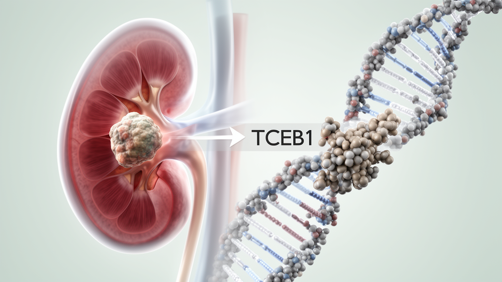
A 52-year-old man with an incidentally discovered kidney tumor was diagnosed with ELOC (TCEB1)-mutant renal cell carcinoma carrying a previously unreported missense variant, c.227A>G (p.Y76C), according to a 2026 case report and literature review. Sequencing also found a truncating TCEB1 variant, c.171_172insCC (p.N58Pfs*48), on the opposite allele. The combination of the tumor’s architecture, immunostaining profile and genetic findings established the diagnosis of a rare molecular subtype first described in 2013 and formally recognized in the 2022 World Health Organization classification. This report concerns one patient, so it adds a molecular observation rather than establishing a new treatment or prognosis.
The patient was admitted to People’s Hospital of Longhui County in China in March 2025 after routine ultrasound revealed a small left renal mass. He had no lumbar pain, chills, fever, urinary urgency, dysuria or gross hematuria, and his routine laboratory findings were unremarkable. Contrast-enhanced computed tomography identified a round, slightly hypodense lesion measuring approximately 14 by 15 mm in the left kidney. Its pre-contrast attenuation was about 35 Hounsfield units, increasing to about 130 units during the arterial phase before it became hypoenhancing relative to the renal parenchyma in the delayed phase. Imaging showed no invasion of the adrenal gland, renal vein or renal sinus, and no extrarenal masses or suspected metastases were detected.
Two days after imaging, the patient underwent retroperitoneal laparoscopic partial nephrectomy. The excised tumor was well circumscribed and measured 1.5 by 1.0 cm; surgical margins were negative, and examination found no involvement of perirenal fat, the renal sinus or lymphatic and blood vessels. Microscopically, the tumor had a nodular architecture divided by smooth-muscle bundles and contained branching tubular and papillary formations supported by delicate fibrovascular cores. The cells were round to oval with sharply defined borders, clear cytoplasm, visible nucleoli, rare mitotic figures and no conspicuous necrosis. Eosinophilic hyaline globules, which have often been described in this subtype, were neither prominent nor identified in the sections available for examination. The pathological stage was pT1a and the International Society of Urological Pathology nucleolar grade was 1. No sarcomatoid or rhabdoid features were present.
Immunohistochemistry supported a renal epithelial neoplasm and helped narrow the diagnosis. Tumor cells diffusely expressed cytokeratin, high-molecular-weight cytokeratin, CK7, CD10, carbonic anhydrase IX, PAX8 and AMACR/P504S, although the P504S staining was weak. The Ki-67 proliferation index was approximately 3%. Vimentin and smooth-muscle actin were absent in the tumor cells but present in the adjacent stroma. TFE3 showed only partial, weak positivity, while CD117, HMB45 and Melan-A were negative. Fluorescence in situ hybridization found no TFE3 gene rearrangement. p53 showed weak-to-moderate staining in roughly 40% of tumor cell nuclei, which the report described as suggestive of wild-type expression. None of these markers is entirely specific for ELOC-mutant renal cell carcinoma, making the interpretation dependent on the combined morphologic, immunophenotypic and molecular findings.
Targeted next-generation sequencing identified four altered loci. The two TCEB1 variants, p.Y76C and p.N58Pfs*48, were found in trans, supporting biallelic inactivation of the gene. The p.Y76C missense change was absent from general population databases and received an AlphaMissense pathogenicity score of 0.9992, above the cited recommended threshold of 0.564. That computational score is a prediction, not functional proof of pathogenicity, but the mutation lies at the interface between Elongin C and pVHL, where other pathogenic variants are concentrated. Sequencing also detected an ABL1 p.A857E alteration with an allelic frequency of about 2.6%, interpreted as a subclonal passenger, and MCL1 amplification, described by the authors as a possible cooperating modifier. No germline variants were detected. Tumor mutational burden was 0.0 mutations per megabase, microsatellite stability was confirmed, and RNA-based fusion testing found no gene rearrangements or actionable genomic alterations.
ELOC, also known as TCEB1, is located at chromosome 8q21 and encodes Elongin C, an essential subunit of the VHL E3 ubiquitin-ligase complex. Under normal oxygen conditions, this complex targets hypoxia-inducible factor alpha for ubiquitin-dependent degradation, limiting hypoxic signaling. Loss-of-function ELOC variants can disrupt the Elongin C–pVHL interface, impairing recruitment and degradation of HIF alpha. Stabilized HIF alpha then enters the nucleus with HIF beta and promotes transcription of oncogenic target genes. Known mutational hotspots include Y79, E92K, A100P, A106D and C112Vfs*3, all within or near the VHL-binding region. Pathogenic ELOC variants are typically accompanied by secondary loss of the other allele, most often through whole-chromosome 8 deletion. In contrast, canonical clear cell renal cell carcinoma drivers such as PBRM1, SETD2, BAP1, TSC1, TSC2, mTOR, PIK3CA, PTEN and ATM are absent in nearly all ELOC-mutant cases reviewed by the report.
The case illustrates why morphology alone is insufficient for identifying this subtype. ELOC-mutant renal cell carcinoma can resemble conventional clear cell renal cell carcinoma, clear cell papillary renal cell tumor and renal cell carcinoma with fibromyomatous stroma. CK7 expression is particularly useful in distinguishing ELOC-mutant tumors from low-grade conventional clear cell carcinoma, while CD10 is usually present in ELOC-mutant tumors and conventional clear cell carcinoma but is generally absent from clear cell papillary tumors. CAIX reflects activity in the HIF pathway, yet its staining pattern must be interpreted with morphology because a cup-shaped pattern is not exclusive to one tumor type. GPNMB and P504S can assist in identifying renal cell carcinoma with fibromyomatous stroma, while TFE3, FH, SDHB and INI1 testing may help exclude other renal tumor categories. When morphology and immunohistochemistry produce an unusual or ambiguous profile, the report argues that genetic testing can clarify the classification.
Most ELOC-mutant renal cell carcinomas are diagnosed at an early pathological stage and low nuclear grade, and their reported clinical course is generally indolent. The literature reviewed for the report describes a male predominance and presentation mainly in middle-aged or older adults. In an 11-patient low-stage series, all patients remained disease-free over a median follow-up of 5.6 years, with two deaths attributed to causes unrelated to renal cell carcinoma. Another reported comparison found superior overall and progression-free survival in patients with ELOC-mutant disease than in patients with stage- and grade-matched VHL-mutant conventional clear cell renal cell carcinoma. These observations come from limited series and cohorts and do not establish the outcome of an individual patient with certainty. Only four stage III cases and two metastatic cases had been documented in the literature considered by the authors. Advanced tumors may acquire additional alterations involving TERT, PMS2 or NF1 and can be associated with higher nuclear grade.
For the reported patient, partial nephrectomy was performed without postoperative adjuvant treatment. He remained alive and without radiographic evidence of recurrence or metastasis through the last reported follow-up on August 20, 2026, 17 months after surgery; imaging performed on August 16 also showed no recurrence. Because the study is a single-patient case report with limited follow-up, its main contribution is the identification and description of a new TCEB1 variant rather than evidence that the variant changes prognosis or treatment response. The authors argue that integrating morphology, immunohistochemistry and sequencing can help distinguish this rare entity from its mimics, avoid unnecessary postoperative adjuvant treatment in surgically resected patients and reduce the risk of distorted clinical-trial data. Evidence for targeted therapies in advanced ELOC-mutant disease remains limited, and further molecularly characterized cases are needed to establish its full biological and clinical spectrum.
Subject of Research: Identification and clinicopathologic characterization of a novel TCEB1 p.Y76C variant in ELOC-mutant renal cell carcinoma
Article Title: Identifying a novel variant in ELOC (TCEB1)-mutant renal cell carcinoma: a case report and literature review
Article References: Wang, Y., Ning, X., Xiao, G., Wu, S., Guo, W., Guo, J., Lei, C., Fan, S., & Ma, X. (2026). Identifying a novel variant in ELOC (TCEB1)-mutant renal cell carcinoma: a case report and literature review. Frontiers in Oncology, 16, Article 1989742. https://doi.org/10.3389/fonc.2026.1989742
Image Credits: AI Generated
DOI: 10.3389/fonc.2026.1989742
Keywords: ELOC, TCEB1, renal cell carcinoma, kidney cancer, p.Y76C, genetic testing, pathology, next-generation sequencing, case report, molecular diagnosis, Identifying, novel
News Source: Ophelia Keating. (October 25, 2026). Case Report Identifies New TCEB1 Variant in Rare Kidney Cancer. Scienmag.