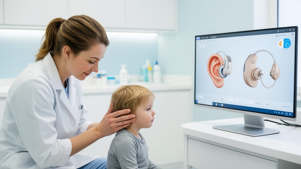
Children undergoing cochlear implant surgery may not need a routine computed tomography scan before their operation, according to a retrospective study from the University Hospital Freiburg in Germany. The research, published in Frontiers in Neurology, compared 164 children who received bilateral cochlear implants between 2018 and 2024 and found that omitting the preoperative temporal bone CT scan did not significantly increase complications or lengthen surgery. Because CT exposes young patients to ionizing radiation, the findings add weight to a growing movement among implant centers toward MRI-only imaging strategies for most pediatric candidates.
Cochlear implants are the standard of care for children with severe to profound sensorineural hearing loss, and timing matters enormously. Newborn hearing screening programs mean that profound hearing loss is often diagnosed shortly after birth, and implantation before speech development is associated with better audiological outcomes, with some children achieving hearing performance comparable to normally hearing peers for up to ten years after implantation. Before surgery, however, every candidate requires careful imaging of the auditory system. MRI excels at showing soft tissue, including the cochlear nerve, retrocochlear and intracranial pathologies, and fluid within the cochlea, while CT provides detailed visualization of the bony anatomy of the temporal bone, including cochlear morphology and structures relevant to surgical planning. Many centers have traditionally ordered both scans, but that combination carries costs: radiation from CT, sedation or anesthesia for MRI in infants, additional healthcare expense, and often a second anesthetic session if the CT is scheduled separately.
The Freiburg team took advantage of a natural experiment within their own department. In 2021, the institution changed its preoperative imaging protocol, moving from routine combined MRI and CT to an MRI-only strategy, with CT reserved for selected cases. Because patient allocation was determined by the timeline of this policy update rather than randomization, the researchers could compare two consecutive eras of practice. They included children implanted bilaterally between 2018 and 2024 who were under ten years old at the time of either simultaneous or second implantation. Ultimately, 105 children received a preoperative CT scan and 59 did not. The children without CT were significantly younger on average, at 21.44 months compared with 26.44 months, a difference the authors attribute to the parallel shift toward earlier implantation as evidence accumulated that operating young is safe and beneficial.
The headline result concerns surgical efficiency and safety. The mean duration of bilateral cochlear implantation was 122.5 minutes without a preoperative CT and 116.1 minutes with one, a difference that was not statistically significant. Total anesthesia time averaged 237.2 minutes without CT versus 226.3 minutes with CT, also not significant in the unadjusted analysis. Importantly, the pattern held regardless of surgical experience: for trainees with fewer than five years of experience, surgery took 184.4 minutes without CT and 159.3 minutes with CT, a non-significant difference, while experienced surgeons took 111.3 and 114.9 minutes respectively. The authors suggest that structured supervision, in which trainees discuss cases with experienced implant surgeons beforehand and can obtain intraoperative guidance, may explain why the absence of a CT did not slow down less practiced hands.
Complication rates were low in both groups. Minor perioperative complications, including hematoma, wound infection, fever, vertigo, pain exacerbation, and cerebrospinal fluid leak, occurred in 10.2 percent of children without a CT scan and 4.8 percent of those with one, a difference that did not reach statistical significance. Major complications and mortality were zero in both cohorts, anesthesiological complications were similar at 6.7 percent versus 8.5 percent, and the median hospital stay was six days in each group. No patient developed postoperative facial nerve palsy, complete or incomplete, and intraoperative X-ray imaging revealed no cases of electrode tip fold-over in either cohort.
The radiological analysis gets to the heart of the debate. Among the 59 children imaged with MRI alone, 83 percent had entirely unremarkable scans, and of the ten children with abnormalities, only one finding, a dysplasia of the vestibule and the inferior and lateral semicircular canals, actually affected surgery, causing resistance during electrode insertion without prolonging operative time. Among the 105 children who also received CT, MRI detected pathological findings in 31.4 percent, of which 23 were considered potentially relevant to implant surgery. Yet only seven of those findings had any impact on the procedure. Intraoperative perilymphatic leakage, known as gusher, occurred in six procedures, five in children with bilateral large vestibular aqueduct and one with bilateral cochlear dysplasia, and one child with malformed apical cochlear turns required an anterior-inferior cochleostomy.
Crucially, the redundancy between the two modalities ran in both directions. Of the potentially relevant findings identified on temporal bone CT, 11 were also detected on MRI and 10 were not, but only a single CT-only finding, the malformed apical cochlear turns, led to a modification of the surgical plan. Conversely, in two patients MRI revealed abnormalities that CT missed: vestibular hypoplasia and a loop of the anterior inferior cerebellar artery contacting the vestibulocochlear nerve. The authors argue that this pattern addresses the central question of the study: the clinical value of routine CT depends on whether it changes surgical management, not merely on whether it detects additional anatomical detail.
The findings do not support abandoning CT altogether. The authors recommend reserving preoperative CT for children in whom clinical or MRI findings suggest relevant bony abnormalities, complex inner ear malformations, or syndromic conditions. In such cases, detailed bony imaging can guide major decisions, such as whether to perform a subtotal petrosectomy for wider exposure and cavity obliteration, and can better delineate the facial nerve course in conditions like CHARGE syndrome or Johanson-Blizzard syndrome. Notably, however, even large vestibular aqueduct and incomplete partition type II, the most frequent abnormalities in this cohort, were detectable on MRI, and one case of incomplete partition type II was identified only on MRI. MRI also remains indispensable for detecting cochlear nerve hypoplasia or aplasia, which directly determines candidacy, and it supports software-assisted surgical planning with cochlear measurements comparable to CT-based evaluation, even though most surgeons remain more familiar with CT-based planning tools such as OTOPLAN.
The study has important limitations that temper the conclusion. It was retrospective and non-randomized, producing unequal cohort sizes and baseline disparities: the children differed in age and in the proportions of sequential versus simultaneous implantation, factors known to influence operative and anesthesia times and complication rates. Although the researchers adjusted for age, residual confounding cannot be excluded, and chronological changes in surgical practice over the 2018 to 2024 study period, including growing experience and different electrode arrays, may have influenced outcomes. The prevalence of inner ear malformations also differed sharply between the CT cohort at 21.9 percent and the MRI-only cohort at 1.7 percent, raising the possibility of selection and detection bias, since MRI is less sensitive than CT for subtle bony abnormalities. Most importantly, the authors caution that a non-significant p-value does not demonstrate equivalence: the wide confidence interval for the complication risk difference, spanning minus 6.3 to plus 14.3 percent, means a true clinical difference cannot be ruled out given the small number of adverse events.
Even so, the study offers a concrete, evidence-based template for centers weighing imaging protocols as pediatric implantation volumes grow and patients get younger. An MRI-based strategy, the authors conclude, may be safe and sufficient in appropriately selected children, reducing unnecessary radiation exposure and healthcare resource use while avoiding redundant diagnostic information, provided CT remains available for the subset of patients with syndromic disorders, complex malformations, or suspicious MRI findings. The team also raises a second radiation question: since routine intraoperative X-rays to check for electrode tip fold-over found no cases, and since electrophysiological methods such as transimpedance matrix measurements may reliably detect fold-over intraoperatively, even that routine imaging step may warrant reconsideration in appropriately selected cases. For now, the message for families and clinicians is one of selective, rather than universal, imaging, with the choice of scans tailored to each child’s anatomy and clinical context rather than applied by default.
Subject of Research: Preoperative CT versus MRI-only imaging before pediatric cochlear implantation
Article Title: Preoperative cranial computed tomography in pediatric cochlear implantation: clinical necessity or optional step?
Article References: Fries, L., Everad, F., Magradze, G., Deffner, F., Aschendorff, A., Arndt, S., & Ketterer, M. C. (2026). Preoperative cranial computed tomography in pediatric cochlear implantation: clinical necessity or optional step?. Frontiers in Neurology, 17, Article 1981222. https://doi.org/10.3389/fneur.2026.1981222
Image Credits: AI Generated
DOI: 10.3389/fneur.2026.1981222
Keywords: cochlear implant, pediatric surgery, preoperative imaging, computed tomography, MRI, temporal bone, ionizing radiation, hearing loss, inner ear malformations, surgical complications, sensorineural hearing loss, large vestibular aqueduct
News Source: Ophelia Keating. (October 24, 2026). Skipping Routine CT Scans Before Children’s Cochlear Implants May Be Safe, Study Finds. Scienmag.