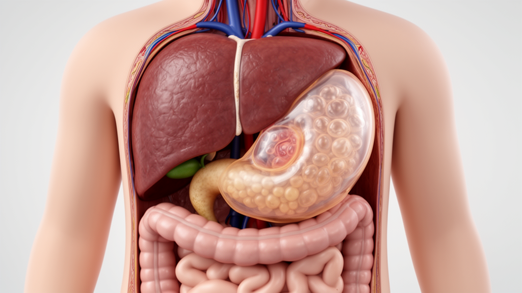
A 14-year-old girl with a large cystic lesion in the upper abdomen was initially diagnosed by radiologists as having a pancreatic pseudocyst, but exploratory surgery identified a hydatid cyst within the lesser sac with posterior extension to the pelvic cavity. The case, reported in BMC Pediatrics , describes an uncommon presentation of hydatid disease in a child and highlights the diagnostic challenge posed by a hydatid cyst that closely resembles a pancreatic fluid collection on imaging. The patient came from a rural livestock-raising region.
She had a one-year history of recurrent left-sided abdominal pain, nausea, and postprandial early satiety, meaning that she developed early fullness after eating. Abdominal ultrasonography and contrast-enhanced computed tomography performed before admission showed a cystic lesion measuring 8.1 by 8.6 cm. The initial radiological interpretation was a pancreatic pseudocyst.
Exploratory laparotomy, conducted through a left upper transverse abdominal incision, instead revealed a large cystic mass within the lesser sac. Posteriorly, it extended downward to the pelvic cavity. The extent of the lesion was therefore established during surgery rather than by the initial imaging report.
Diagnostic aspiration of the mass yielded clear to slightly yellowish fluid, leading the surgical team to suspect hydatid disease. The infection is caused by the parasitic zoonotic organism Echinococcus granulosus and most commonly affects the liver and lungs, while retroperitoneal involvement is rare in children. The aspiration finding supported reconsideration of that diagnosis, and subsequent treatment was directed at the hydatid cyst.
The surgeons used 70% ethyl alcohol as a scolicidal agent, followed by partial pericystectomy and evacuation of the cyst contents. The residual cavity was then irrigated with povidone-iodine and normal saline. Two drains were placed: one within the cyst cavity and another in the adjacent residual cavity for external drainage.
No intraoperative cyst rupture, fluid leakage, or spillage of cystic contents occurred during the operation. In the absence of these events, the patient received adjunctive antihelminthic treatment with albendazole at 10 mg/kg/day for 21 days. At six months after the operation, the patient remained asymptomatic and had no evidence of postoperative complications.
This was a single-patient case report rather than a comparative study, so it cannot establish the frequency of retroperitoneal hydatid disease in children or determine how reliably such cysts can be distinguished from pancreatic pseudocysts. Its reported value is instead to document a rare anatomical presentation in a symptomatic adolescent and to show that hydatid disease should be considered in the differential diagnosis of upper-abdominal cystic masses in children from endemic or livestock-raising regions, even when imaging suggests a pancreatic pseudocyst.
Subject of Research: A pediatric hydatid cyst of the lesser sac with retroperitoneal extension initially mistaken for a pancreatic pseudocyst.
Article Title: Hydatid cyst of the lesser sac with retroperitoneal extension mimicking a pancreatic pseudocyst in a child: a case report
Article References: Hakimi, T., Shirpor, A. S., Rahmani, F. R., & Jawed, M. A. (2026). Hydatid cyst of the lesser sac with retroperitoneal extension mimicking a pancreatic pseudocyst in a child: a case report. BMC Pediatrics. https://doi.org/10.1186/s12887-026-07878-0
Image Credits: AI Generated
DOI: 10.1186/s12887-026-07878-0
Keywords: pediatric hydatid disease, Echinococcus granulosus, retroperitoneal cyst, lesser sac, pancreatic pseudocyst, abdominal imaging, pediatric surgery, albendazole, case report, cystic mass, Hydatid, cyst
News Source: Ophelia Keating. (October 24, 2026). Pediatric hydatid cyst mimicked a pancreatic pseudocyst in case report. Scienmag.