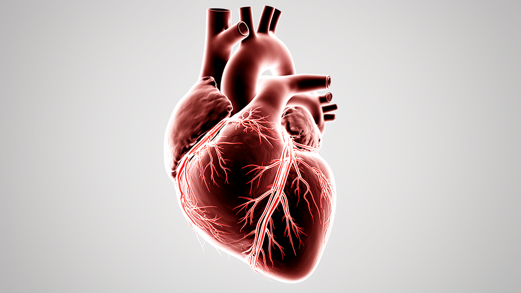
A perfusion pressure of 60 mmHg was the lowest at which the coronary arteries showed complete and homogeneous opacification with minimal perfusion-related artefacts in all evaluable cases of a pilot study on postmortem computed tomography angiography, published in the International Journal of Legal Medicine. The exploratory investigation, led by Ngoc Huyen-Chau Feldmann and colleagues, examined three intravascular pressure groups of 40, 60 and 80 mmHg in twelve postmortem cases, of which six yielded analysable imaging data. Using a modified contrast agent pump with coupled real-time pressure monitoring and control, the researchers found that 40 mmHg produced discontinuous, segmental contrast filling of the coronary arteries, whereas 60 and 80 mmHg achieved continuous opacification of the main coronary stems and their smaller branches.
In one case examined at 80 mmHg, the peripheral coronary branches were depicted more completely than at 60 mmHg, an observation the authors explicitly state requires confirmation in a larger cohort. The findings may contribute to the methodological standardisation of pressure-based postmortem angiography protocols in forensic practice. Coronary artery disease continues to rank among the leading causes of mortality worldwide and represents a major underlying factor in cases of sudden cardiac death, making accurate postmortem assessment of the coronary arteries essential for determining the cause and mechanism of death.
Postmortem CT angiography was introduced in 2008, when Grabherr and colleagues presented an approach to simulate an artificial circulation using a specifically developed perfusion device, enabling both arterial and venous imaging phases. A protocol defining perfusion parameters such as flow rate and contrast agent volume followed in 2011 to ensure high-quality visualisation and comparability, and the advantages of the method for postmortem examinations have since been demonstrated in multiple studies. Several investigations have shown that coronary postmortem CT angiography agrees well with autopsy and, in selected settings, histopathology in detecting relevant coronary pathology, including stenoses, occlusions, bypass grafts and stents.
However, studies focusing on quantitative cardiovascular measurements have reported only limited correlation with autopsy findings, and recent review articles emphasise that in deaths from presumed natural cardiac causes, postmortem imaging currently remains less accurate than conventional autopsy and should primarily be regarded as a complementary diagnostic tool rather than a replacement. The technical and interpretive challenges of coronary imaging differ considerably between living patients and the postmortem setting. In clinical CT angiography, cardiac motion, heart rate variability and the small calibre of distal branches can impair coronary visualisation.
In the deceased, vascular collapse, increased blood viscosity and the absence of active circulation may lead to inhomogeneous contrast filling, discontinuous segmental opacification patterns, or contrast pooling that can mimic or obscure clinically relevant lesions. In the present work, the term beads-on-a-string refers to a discontinuous, segmental pattern of contrast opacification caused by incomplete perfusion, and not to a vascular pathology such as fibromuscular dysplasia. Such technique-related artefacts of multi-phase postmortem CT angiography have been described systematically, and artefacts may lead to false-positive interpretations of coronary stenosis if perfusion conditions are not adequately controlled.
A further complication is that the contrast agent mixtures used in postmortem angiography are oil based and considerably more viscous than the water-based media used in living patients, which together with the absence of active circulation means that pressure values established in clinical angiography cannot be transferred directly to the postmortem setting. One parameter that had not been sufficiently investigated was the intravascular pressure during the contrast agent injection and its effect on image quality. Previously reported pressure values in postmortem angiography most likely referred either to the injection pressure of the contrast pump or to the pressure measured at the femoral artery access site, rather than to the intravascular pressure within the coronary circulation.
In 2024, Fischer published a method enabling the continuous recording of intravascular contrast agent pressure during postmortem CT angiography using the Contrast Agent Application Device system with real-time pressure sensors and radial artery catheterisation, and the present study is based on that method. The system comprises a roller pump with a variable flow rate of 100 to 1200 mL/min, electromagnetic tubing clamps for contrast flow regulation, pressure sensors covering a range of 0 to 200 mmHg calibrated per DIN EN 60770, and software-based control for pressure- or volume-regulated operation. The software adjusted the pump output in real time to maintain the predefined target pressure within a tolerance of plus or minus 5 mmHg during both arterial and venous perfusion phases, and pressure data were recorded throughout the procedure.
The study examined twelve cases, nine male and three female, with an average age of 58 years and a range of 26 to 86 years. Inclusion criteria consisted of a short postmortem interval of one to three days, absence of externally evident traumatic injuries, and no visible signs of decomposition, while cases involving suspected homicidal death, suicide, or tissue donation were excluded. None of the cases originated from a hospital setting, and none had a documented history of extensive fluid therapy or resuscitation.
The twelve cases were allocated to the three pressure groups, and two evaluable cases per group were available for analysis. In the remaining six cases, adequate image evaluation was not possible due to technical complications, including massive atherosclerosis of the femoral artery, catheter dislocation and pre-existing vascular pathologies such as rupture of the aorta. Each analysable pressure group therefore included two cases with complete datasets, reflecting the exploratory nature of the study.
Image interpretation was carried out as a consensus reading by two board-certified forensic pathologists experienced in forensic radiology, with 22 and 10 years of experience, and a board-certified radiologist with 14 years of experience, who were not blinded to the pressure group. The imaging protocol began with a native whole-body CT scan on a GE Optima 660 64-row multidetector scanner, which provides the non-enhanced images required for comparison during interpretation, for example for the differentiation of calcifications from the contrast agent mixture; the authors note it was not used as a pre-screening to exclude cases with relevant vascular pathology before catheterisation, which they describe as a methodological refinement for future studies. The femoral artery and vein were then prepared, and an 18 French arterial catheter and a 16 French venous catheter were placed using the Seldinger technique.
A small 3 French arterial catheter was placed in the radial artery for pressure monitoring, chosen because femoral arterial measurements had been shown to be substantially influenced by pump-related pulsatility, whereas radial artery monitoring yielded more stable and reliable measurements. Venous pressure monitoring data were not included in the analysis. The contrast agent mixture was 4 percent Angiofil dissolved in rapeseed oil, whose kinematic viscosity of 74 mm²/s at 20 °C is comparable to Paraffinum liquidum and higher than Paraffinum perliquidum.
No dynamic phase was performed, in order to isolate the effect of a single, continuously controlled static pressure. The results revealed a clear pattern across the pressure groups. At 40 mmHg, the target pressure was reached after the administration of 254 mL and 325 mL of contrast agent, but the left anterior descending, left circumflex and right coronary arteries were only partially opacified in their central portions, with incomplete visualisation towards the distal segments and the cardiac apex.
Smaller branches, including septal branches arising from the left anterior descending artery, were inconsistently visualised, and contrast enhancement appeared discontinuous, showing the characteristic beads-on-a-string pattern. At 60 mmHg, reached after 550 mL and 629 mL, contrast was continuously distributed throughout the coronary arteries, with the main stems opacified to the distal segments and the cardiac apex, smaller septal and diagonal branches well visualised along their entire course, and the sinoatrial nodal artery fully opacified in all examined specimens. At 80 mmHg, reached after 423 mL and 1091 mL, the vessels remained fully opacified along their entire course including peripheral branches, with contrast distributed homogeneously in both central and peripheral vascular structures.
The volume required to reach the target pressure varied considerably between cases and depended on the individual vascular status, in particular vascular compliance and pre-existing vascular pathology, rather than on the target pressure alone. The pressure levels were chosen based on the assumption that, in intensive care settings, a mean arterial pressure of 60 mmHg is considered sufficient for organ perfusion, with a lower and a higher pressure evaluated additionally so that perfusion conditions approximate physiological values as far as the postmortem setting allows. The authors argue that this matters because conventional autopsy can assess the main coronary arteries morphologically in detail, but interseptal branches and other small branches are difficult or even impossible to dissect, and the functional aspect of blood flow through a narrowed coronary lumen cannot be evaluated by dissection alone.
The observations suggest that perfusion pressure may be a further technical factor associated with image quality in postmortem angiography, alongside previously identified sources of pseudo-pathological findings such as gas bubbles, thrombus formation, contrast layering and vessel collapse. Because perfusion pressure can be adjusted during the procedure, it may help to reduce such artefacts. More homogeneous opacification might also facilitate the assessment of microvascular alterations and coronary anatomical variants, including aneurysms, dissections, and congenital or acquired anomalies, although the authors caution that this could not be verified in the present study because no direct autopsy or histopathological reference standard was used for that purpose.
The limitations of the pilot study are substantial and are stated candidly by the authors. The exclusive focus on the coronary arteries means the observations may not be directly transferable to other vascular compartments or the venous phase, since postmortem haemodynamics can vary substantially between different vascular territories. Neither pressure has been tested against the cerebral circulation, so it cannot be assumed that a pressure found favourable for coronary opacification is equally suitable there; protocol development studies have instead used the cerebral arteries as the main criterion for selecting injection pressure and volume because their aim was whole-body perfusion quality.
Due to the small sample size of twelve cases, of which six were evaluable, no statistical analysis was a primary intention, and a larger sample is required for further statistical evaluation. No direct correlation with macroscopic autopsy or histopathological reference standards was performed, such validation is still lacking for postmortem imaging of cardiac deaths in general, and diagnostic accuracy was not assessed. The readers were not blinded to the pressure group, and a blinded, independent scoring design would be appropriate for a follow-up study.
The authors conclude that 60 mmHg provided sufficient contrast opacification for diagnostic visualisation in the two evaluable cases studied at that pressure, but that this should not be regarded as a generally established threshold and requires confirmation in larger, adequately powered cohorts before the pressure values reported here can be considered validated.
Subject of Research: Effect of perfusion pressure on coronary artery image quality in postmortem CT angiography
Article Title: Influence of perfusion pressure on image quality in postmortem CT angiography (PMCTA) of coronary arteries: a pilot study
Article References: Feldmann, N. H.-C., Sabel, B. O., Bruch, G. M., & Fischer, F. T. (2026). Influence of perfusion pressure on image quality in postmortem CT angiography (PMCTA) of coronary arteries: a pilot study. International Journal of Legal Medicine. https://doi.org/10.1007/s00414-026-04050-2
Image Credits: AI Generated
DOI: 10.1007/s00414-026-04050-2
Keywords: postmortem CT angiography, PMCTA, coronary arteries, perfusion pressure, forensic radiology, sudden cardiac death, contrast agent, pilot study, Influence, perfusion, pressure, image
News Source: Ophelia Keating. (October 24, 2026). Research report: Influence of perfusion pressure on image quality in postmortem CT angiography (PMCTA) of coronary arteries: a pilot study. Scienmag.