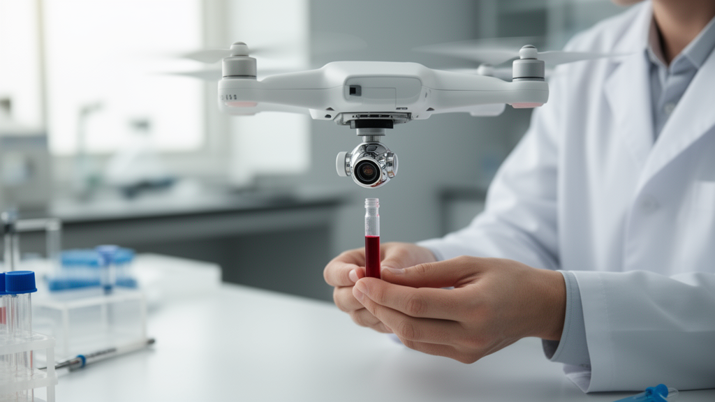
A lightweight drone fitted with a gold-nanoparticle biosensor detected dengue virus antigen in human serum with a reported limit of detection of 0.91 micrograms per milliliter and a visible test-line signal within five seconds. The prototype combines a lateral-flow assay, a compact unmanned aerial vehicle, image analysis based on YOLOv5 and a QR-code-linked reporting system. Researchers characterized the sensor in laboratory tests and limited field conditions, rather than demonstrating autonomous surveillance during a dengue outbreak. In the assay, dengue antigen separated a DNA aptamer from its gold-nanoparticle conjugate, allowing the released nanoparticles to bind immobilized lysozyme and produce a red band visible to the naked eye.
The study was designed as a proof-of-concept for decentralized dengue screening. Its central idea is to carry a small diagnostic strip to a location that can be reached by drone, perform the test near the collection point and transmit the result through a digital workflow. The platform used a DJI Mini 3 Pro weighing less than 249 grams, with dimensions of 251 by 362 by 70 millimetres, an intelligent-flight battery capacity of 2,453 milliampere-hours and a stated top speed of 16 metres per second in S mode under sea-level, no-wind conditions. The lateral-flow strip was attached to the drone with a double-sided adhesive and enclosed in translucent Plexiglas to protect it during flight. A QR code placed on the aircraft opened a reporting webpage through which a user could transmit results to laboratory personnel.
At the biochemical core of the system is a 34-base DNA aptamer designed to recognize polyvalent dengue antigen. Researchers synthesized gold nanoparticles by reducing chloroauric acid with trisodium citrate, then mixed 24 microlitres of aptamer with 500 microlitres of the nanoparticle suspension at a pH of 9.2 to 9.6. After incubation, bovine serum albumin was added as a blocking agent, and the conjugate was washed, resuspended in phosphate-buffered saline and stored at 4 degrees Celsius. Zeta-potential analysis gave the unmodified particles a mean surface charge of minus 37.56 millivolts, consistent with strong electrostatic repulsion and colloidal stability. Dynamic light scattering measured an average hydrodynamic diameter of 11.59 nanometres, while transmission electron microscopy gave a particle diameter of 12.43 plus or minus 0.20 nanometres. The particles were well dispersed and spherical.
Aptamer attachment altered the nanoparticles in ways that were consistent with successful functionalization. Their surface-plasmon-resonance band shifted from 521 to 526 nanometres after conjugation, accompanied by a decrease in absorbance, indicating a change in the local environment around the gold surface. Dynamic light scattering also recorded an increase in hydrodynamic diameter to 17.11 nanometres, which the authors attributed to the aptamer coating rather than aggregation. The investigators characterized the interaction between the aptamer and dengue antigen through molecular docking and an electrophoretic mobility shift assay. Docking produced a score of minus 326.89, which the authors interpreted as strong predicted binding affinity, although a ligand root-mean-square deviation of 66.33 angstroms indicated considerable conformational flexibility or imperfect structural alignment after docking. In the mobility assay, the conjugate shifted relative to the free aptamer, as did the aptamer–antigen complex, supporting the formation of the intended molecular complexes.
The lateral-flow sensor used a sample pad, conjugate pad, nitrocellulose membrane and absorbent pad mounted in a plastic cassette. A 70-microlitre sample was applied to the pad and moved by capillary action. When dengue antigen was present, it bound the aptamer and promoted release of the gold nanoparticles from the complex. The free nanoparticles then reached a test line containing immobilized lysozyme and formed a visible red signal. In the absence of dengue antigen, the gold–aptamer complex remained associated and did not produce the test-line band. During a separate verification using 80-microlitre samples, dengue antigen and phosphate-buffered saline controls were run at room temperature for 30 seconds. Of three tested stages—free nanoparticles, gold–aptamer conjugate and gold–aptamer–dengue antigen complex—only the complete three-component stage generated a signal. Scanning electron microscopy showed the porous and fibrous structures of the strip components and the deposition of biomaterial on the test line.
The researchers optimized the pH and aptamer loading before evaluating analytical performance. The gold nanoparticles performed well at pH 7, while increasing the amount of aptamer initially strengthened the test-line signal before reaching a plateau or modest decline at higher loadings. The investigators attributed that downturn to steric hindrance or excessive surface capping, which could impede the release of nanoparticles required by the competitive assay format. Strips were then tested across dengue-antigen concentrations from 1 to 100 micrograms per milliliter. Test-band intensity rose with concentration, and ultraviolet-visible spectroscopy was used to examine the associated changes in nanoparticle dispersion. Densitometric analysis with ImageJ produced a calibration coefficient of determination of 0.994 within the working range, allowing the authors to describe the assay as suitable for semiquantitative antigen measurement. Each concentration was tested in seven replicates for the intensity analysis.
The reported limit of detection was calculated as three times the standard deviation of the response divided by the slope of the calibration curve, using seven observations. It was 0.91 micrograms per milliliter, and the study’s highlights state that a visible result could be obtained within five seconds. Recovery tests produced values of 92.79% and 105.96%, with relative standard deviations of 1.61% and 2.33%. Student’s t-values of 0.850 and 0.442 were below the critical value of 2.447 for six degrees of freedom at a two-tailed 0.05 significance level. In separate repeatability measurements, the reported intra-day mean was 112.41% plus or minus 1.18%, with an RSD of 1.05%, while the inter-day mean was 113.32% plus or minus 1.45%, with an RSD of 1.28%. These are analytical assay results rather than measurements of accuracy against a broad clinical population.
Selectivity experiments tested chikungunya virus and hepatitis C virus at 1 microgram per milliliter, the lowest dengue-antigen concentration used in the reported comparison. Neither produced a test-line signal, supporting specificity for the intended target under the conditions tested. The researchers also evaluated three independently collected human-serum samples, each tested in triplicate, and observed dengue signals in spiked serum. A commercial gold-standard lateral-flow kit was used for comparison, although the supplied article does not provide a detailed clinical agreement table. The human-serum work was approved by the Jamia Hamdard Institutional Ethics Committee, and participants provided written informed consent. The study nevertheless involved a small set of specimens, so the serum results support feasibility rather than establishing performance across diverse patients or a validated clinical diagnosis.
Automated image analysis was added to reduce dependence on subjective visual interpretation. The YOLOv5 small model was used first to locate the predefined result window, not to diagnose patients. On a 47-image working set, the region-of-interest localization achieved a mean average precision at an intersection-over-union threshold of 0.5 of about 0.995 and a value across thresholds from 0.5 to 0.95 of about 0.945. A separate linear support-vector-machine classifier assessed positive or negative strips. Its 25-image test set contained 18 positive and seven negative images, producing six true negatives, one false positive, no false negatives and 18 true positives. Reported accuracy was 96.00%, sensitivity 100.00%, specificity 85.71%, precision 94.74% and F1 score 97.30%, with a reported ROC-AUC and average precision of 1.000. The authors cautioned that the reconstructed image dataset was small and that historical independence of the training and test sets could not be guaranteed, making these results preliminary model-development performance rather than independent clinical validation.
The reporting layer, called the Delhi Against Dengue platform, links a QR code to a mobile webpage showing the result, a unique barcode for an electronic prescription and summary counts of positive and negative reports. It also presents dengue information and graphs of reported trends from 2020 to 2024. This digital element is designed to support communication with clinicians and laboratory authorities, but the platform’s surveillance value was not established through a prospective outbreak trial. The authors identify several constraints to operational use: drone flight endurance of roughly 20 to 30 minutes, payload limits, adverse-weather sensitivity, communication range and bandwidth, crash risk, and aviation, privacy and security requirements in populated or sensitive areas. Large multicenter clinical studies, environmental robustness tests, field trials, regulatory clearance and optimized hardware would be needed before deployment. The demonstrated result is therefore a technically characterized drone-integrated diagnostic prototype, while autonomous outbreak surveillance, real-time epidemiological mapping and large-scale remote deployment remain future objectives.
Subject of Research: Proof-of-concept drone-integrated lateral-flow biosensor for dengue antigen detection
Article Title: Intelligent drone-based biosensor for the detection of dengue virus antigen towards smart unmanned aerial-vehicle platform-based virus diagnosis
Article References: Hasan, M. R., Khan, S., Naithani, D., Sharma, P., Mehta, Y., Ansari, S., kumar, S., Naikoo, U. M., Baliyan, A., Abbas, M., Parayangat, M., Kalathil, S., & Narang, J. (2026). Intelligent drone-based biosensor for the detection of dengue virus antigen towards smart unmanned aerial-vehicle platform-based virus diagnosis. Frontiers in Bioengineering and Biotechnology, 14, Article 1907197. https://doi.org/10.3389/fbioe.2026.1907197
Image Credits: AI Generated
DOI: 10.3389/fbioe.2026.1907197
Keywords: dengue virus, biosensor, lateral-flow assay, gold nanoparticles, DNA aptamer, unmanned aerial vehicle, YOLOv5, point-of-need testing, digital reporting, Intelligent, drone-based, detection
News Source: Kristina Jarvis. (October 21, 2026). Drone-integrated biosensor detects dengue antigen in five seconds. Scienmag.