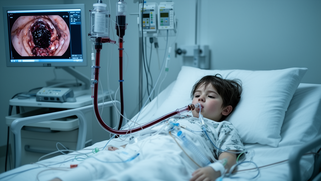
A previously healthy 7-year-old girl survived group A streptococcal toxic shock syndrome complicated by acute necrotizing tracheobronchitis, severe lung injury and multiorgan dysfunction, according to a 2026 case report. The child required veno-venous extracorporeal membrane oxygenation, continuous renal replacement therapy, broad antimicrobial and immunomodulatory treatment, and repeated bronchoscopy before recovering airway function. She was discharged after 27 days, although a later tracheal granulation lesion required endoscopic removal. The report emphasizes that bronchoscopy documented the progression from airway inflammation to extensive erosive necrosis and pseudomembrane formation, but the single-patient evidence cannot establish why the airway was damaged or which treatment produced recovery.
The girl was admitted to a pediatric intensive care unit in Jinhua, China, after six days of fever and three hours of chest tightness and dyspnea. On arrival she was in septic shock with a blood pressure of 82/55 mmHg, a heart rate of 167 beats per minute, marked respiratory distress, cyanosis, cool extremities, delayed capillary refill, and widespread petechiae, purpura and ecchymoses. Coagulation abnormalities met the reported overt disseminated intravascular coagulation score. She was intubated immediately and treated with fluid resuscitation, norepinephrine, linezolid, vancomycin and meropenem. Ventilation was difficult because of pulmonary edema, poor compliance, abundant frothy secretions and high airway resistance, with delivered tidal volumes of 40–50 mL, equivalent to 2.1–2.6 mL/kg.
Before extracorporeal support, arterial blood gas testing at an inspired oxygen fraction of 1.0 showed profound respiratory and circulatory compromise: pH was 7.11, arterial carbon dioxide pressure 53.2 mmHg, arterial oxygen pressure 27.8 mmHg, lactate 7.4 mmol/L and base excess −12.4 mmol/L. At a mean airway pressure of 35 cmH₂O, the oxygenation index was 125.9. Veno-venous ECMO was established at 04:38 on hospital day 2 for hypoxemia that persisted despite conventional ventilation. Blood flow and sweep gas flow were each approximately 2 L/min, with blood flow equivalent to about 105 mL/kg/min, and heparin was titrated to an activated clotting time of 160–180 seconds. Continuous renal replacement therapy was also started for oliguric acute kidney injury and fluid overload.
Laboratory findings on day 2 indicated profound hematologic, renal, hepatic and cardiac dysfunction. The white blood cell count was 0.52 × 10⁹/L, absolute neutrophil count 0.20 × 10⁹/L and platelet count 26 × 10⁹/L. International normalized ratio was 3.55, activated partial thromboplastin time 150.1 seconds, fibrinogen 1.96 g/L and D-dimer 28,870 µg/L. Creatinine was 167.5 µmol/L, aspartate aminotransferase 638 U/L and troponin I exceeded 50 ng/mL. The first 24-hour fluid balance was 4,030 mL in versus 490 mL out, with urine output of 0.48 mL/kg/hour. The child also had severe pneumonia and acute respiratory distress syndrome, placing the illness within a syndrome of acute multiorgan dysfunction.
Conventional blood and sputum cultures were negative after 48 hours, and blood cultures also remained negative at five days. Metagenomic next-generation sequencing of blood and bronchoalveolar lavage fluid, performed on day 2 with a reported turnaround of about 24 hours, instead detected Streptococcus pyogenes in both specimens and Mycoplasma pneumoniae in the lavage fluid. Gram staining of a respiratory specimen showed numerous neutrophils with gram-positive cocci in pairs and short chains. Respiratory-virus and SARS-CoV-2 tests were negative. The authors considered the group A streptococcus the primary pathogen because it was detected in blood, where M. pneumoniae was absent, and the shock, purpura and disseminated intravascular coagulation matched streptococcal toxic shock. They nevertheless classified the case as probable rather than culture-confirmed because the organism was identified by sequencing rather than isolation.
Serial fiberoptic bronchoscopy revealed a rapidly destructive airway process. On day 2, the carina was sharp, but the bronchi were congested and edematous and contained viscous secretions. By day 4, the mucosa was hyperemic and bleeding with purulent material. During ECMO support on day 7, erosive necrosis extended throughout the tracheobronchial tree, with yellow, moss-like pseudomembranes and blood staining. Examination on day 9 again showed marked erosive necrosis and old hemorrhage in the tracheal and bronchial mucosa. These observations established acute necrotizing tracheobronchitis, a rare complication that can allow sloughed membrane or debris to obstruct the airway. No acute-phase airway histology was obtained, and the bronchoscopic findings were available from procedure notes rather than retained images.
The clinical team used multiple supportive and disease-directed interventions, but their relative effects cannot be separated. Antimicrobial treatment continued unchanged after sequencing identified S. pyogenes, rather than being narrowed to the preferred penicillin-and-clindamycin combination for severe invasive group A streptococcal disease. Vancomycin troughs measured 46.9 and 32.9 µg/mL during CRRT, both above the reported target of 10–20 µg/mL. Methylprednisolone was begun at approximately 2 mg/kg every 12 hours and briefly increased to approximately 10 mg/kg per dose every 12 hours before being reduced. IVIG was prescribed at 10 g per dose, and tocilizumab at 240 mg, or approximately 12.6 mg/kg per dose. Hemoperfusion was performed twice, and ulinastatin, sivelestat, albumin, blood products, mannitol, milrinone and norepinephrine were also given. Important details, including administration dates and numbers of doses for IVIG and tocilizumab, antibiotic stop dates and several vasoactive doses, were absent from the records.
Cardiac and lung imaging helped characterize the illness. Lung ultrasound obtained on day 3 showed uneven aeration, with confluent B-lines in some regions and A-lines in others, consistent with the extensive pulmonary involvement seen during ECMO support. A progress note on day 4 recorded a left ventricular ejection fraction of 62% and mild tricuspid regurgitation. The preserved systolic function did not fit the profound ventricular failure usually associated with veno-arterial ECMO, and the clinicians therefore favored sepsis-associated myocardial injury over fulminant myocarditis despite the markedly elevated troponin. Peak interleukin-6 concentration was 942,990 pg/mL and peak procalcitonin exceeded 100 ng/mL, indicating a severe inflammatory and infectious response, although the tocilizumab and IVIG exposure could not be reconstructed fully from the chart.
Recovery proceeded as the airway findings improved. ECMO was discontinued and the cannulas removed on day 9, after review by the vascular surgery team, and CRRT ended on day 10. On day 13, bronchoscopy showed that both main bronchi were patent, the mucosa was pale and only focal erosion remained above the carina; the child was extubated that day and initially supported with high-flow nasal cannula. She transferred to the ward on day 16, stopped supplemental oxygen on day 19 and was discharged on hospital day 28 while alert, afebrile and breathing comfortably. Adverse events included supratherapeutic vancomycin concentrations, Candida detected in urine and stool after prolonged antibiotics and high-dose corticosteroids, and weakness that improved from MRC grade 3 to grade 4 before discharge.
About three months after discharge, persistent throat phlegm and an intratracheal lesion led to another admission. Bronchoscopy found mild scar-like narrowing in the mid-trachea and a polypoid lesion in the lower trachea, which was removed with an electrosurgical snare. Histology showed granulation tissue with hemorrhage and hemosiderin but no malignancy, consistent with post-inflammatory repair. A subsequent admission for bronchopneumonia and a Klebsiella pneumoniae urinary tract infection resolved with antibiotics. At the most recent contact, her guardians reported normal peer interaction, counting and arithmetic, and no need for home oxygen, but standardized cognitive testing, formal spirometry and surveillance bronchoscopy had not been performed. The report therefore provides a detailed account of a rare airway complication and a multidisciplinary recovery, while its culture-negative diagnosis, incomplete treatment documentation and lack of a comparison group limit conclusions about causation and generalizability.
Subject of Research: A case of pediatric group A streptococcal toxic shock syndrome with acute necrotizing tracheobronchitis during veno-venous ECMO.
Article Title: Case Report: Pediatric group A streptococcal toxic shock syndrome complicated by acute necrotizing tracheobronchitis requiring veno-venous extracorporeal membrane oxygenation
Article References: Deng, H., Chen, K., & Chen, K. (2026). Case Report: Pediatric group A streptococcal toxic shock syndrome complicated by acute necrotizing tracheobronchitis requiring veno-venous extracorporeal membrane oxygenation. Frontiers in Pediatrics, 14, Article 1900220. https://doi.org/10.3389/fped.2026.1900220
Image Credits: AI Generated
DOI: 10.3389/fped.2026.1900220
Keywords: pediatric toxic shock, group A Streptococcus, necrotizing tracheobronchitis, extracorporeal membrane oxygenation, metagenomic sequencing, bronchoscopy, acute respiratory distress syndrome, multiorgan dysfunction, pediatric intensive care, Case, Report, Pediatric
News Source: Ophelia Keating. (October 21, 2026). Child with strep toxic shock develops necrotic airways during ECMO. Scienmag.