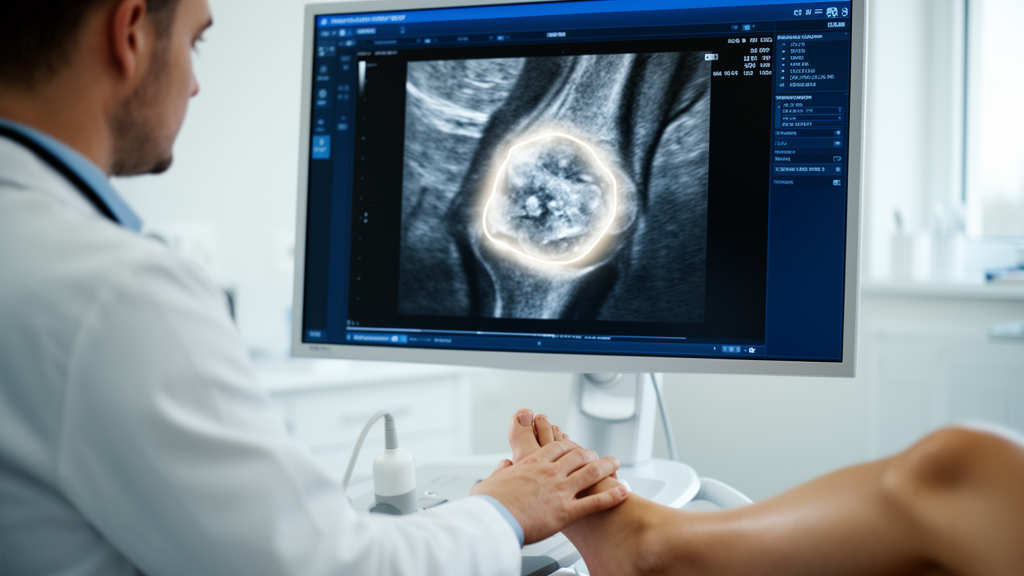
For millions of people living with gout, the painful reality of the disease is written in lumps of crystallized uric acid called tophi. These chalky deposits build up slowly around joints, tendons and soft tissues, and their size and number are among the most important clues doctors use to judge how well treatment is working. Now a team of researchers in Beijing has shown that artificial intelligence can take over one of the most tedious steps in tracking them: outlining tophi, pixel by pixel, on ultrasound images. The study, published in BMC Medical Imaging, demonstrates that four different deep neural networks can automatically segment tophi with an accuracy that rivals experienced sonographers, opening the door to fast, objective and standardized measurement of a lesion that has long resisted quantification.
Gout is one of the most common inflammatory joint diseases worldwide, driven by elevated uric acid that crystallizes into monosodium urate deposits. When these deposits accumulate into visible, palpable masses, they are called tophi, and their presence marks a more advanced stage of disease. Clinical guidelines from organizations such as the American College of Rheumatology and the European League Against Rheumatism emphasize that monitoring tophi is central to managing gout, because shrinking tophi is direct evidence that urate-lowering therapy is working. But measuring them has always been awkward. Physical examination is subjective and struggles with small or deeply seated deposits, while dual-energy CT and MRI, though accurate, are expensive and expose patients to radiation or logistical burdens.
Musculoskeletal ultrasound has emerged as the practical middle ground. It is widely available, radiation-free and sensitive to the characteristic hyperechoic appearance of urate crystals. Yet ultrasound has its own weaknesses. The image quality depends heavily on the skill of the operator, the borders of a tophus can blur into surrounding tissue, and atypical sonographic features can fool even trained eyes. Worse still, if researchers want to compute tophus volume or track change over time, someone must laboriously trace the outline of each lesion by hand on every image. That manual delineation is slow, inconsistent between observers, and simply not scalable to large clinics or clinical trials. The new study set out to test whether deep learning could remove this bottleneck.
The researchers assembled a dataset of 730 original ultrasound images showing tophi, collected at Peking Union Medical College Hospital and its collaborators. The images were randomly divided at the patient level into a training set of 558 images, a validation set of 89 images and an internal test set of 83 images, a split designed to prevent any single patient’s scans from appearing in both training and testing, which would artificially inflate performance. To probe how well the models might generalize beyond their home institution, the team also compiled an independent external test set of 30 images. Four well-established segmentation architectures were then trained to trace tophi automatically: U-Net, Attention U-Net, TransUNet and Deeplabv3+.
Each of these networks represents a different strategy for teaching a machine to see. U-Net, the workhorse of medical image segmentation, uses an encoder-decoder structure that compresses an image into abstract features and then expands it back to full resolution, learning fine boundaries along the way. Attention U-Net adds attention gates that help the network focus on relevant regions and suppress background noise. TransUNet injects transformer blocks with multi-head self-attention into the U-Net framework, allowing it to capture long-range context across the image rather than just local patterns. Deeplabv3+ relies on atrous spatial pyramid pooling, a technique that probes the image at multiple scales simultaneously to capture objects of varying size. All four were trained with binary cross-entropy loss to produce a pixel-level mask of the tophus.
Performance was judged with two standard overlap metrics: the Jaccard Similarity Coefficient, which measures the intersection of the predicted and true regions divided by their union, and the Dice Similarity Coefficient, which weights the overlap differently and is generally a bit more forgiving. On the internal test set, Deeplabv3+ achieved the best average JSC of 64.60 percent and average DSC of 76.77 percent, with TransUNet close behind at 63.59 percent JSC and 76.29 percent DSC. U-Net, while slightly less accurate, was by far the leanest model, with only 31.00 million parameters and a per-image inference time of 68.78 milliseconds, fast enough for real-time use at the bedside. All four models converged smoothly during training and validation, and none showed signs of overfitting.
When the models faced the external test set of images they had never seen from a different distribution, performance dipped slightly, as is typical for medical AI, but the drop was modest and no statistically significant differences emerged between the four architectures. More striking was the comparison against human experts. The segmentation accuracy of Deeplabv3+ and TransUNet was comparable to that of senior sonographers and clearly superior to that of junior ones. In other words, the best models performed at the level of experienced specialists, and outperformed less experienced operators, suggesting the technology could help level the playing field in settings where expertise is scarce.
Accuracy alone is not enough for a clinical tool; a model must also avoid hallucinating tophi where none exist. To test this, the researchers ran all four networks on ultrasound images from gout patients that contained no tophi, including images showing other non-tophaceous lesions. All models generated only very small segmented regions, with overall low false positive rates. TransUNet proved the most conservative, showing the lowest false positive rate across most non-tophaceous lesion types. This specificity matters because a system that flags normal tissue as disease would erode clinician trust and could lead to inappropriate treatment decisions.
The implications reach beyond convenience. Because the models produce precise pixel-level outlines, they enable quantitative assessment of tophus volume, something manual measurement has never been able to deliver reliably at scale. A validated segmentation engine could be embedded into ultrasound machines to give clinicians instant, reproducible measurements during a scan, or used retrospectively to analyze thousands of images in clinical trials of new urate-lowering drugs. Standardized, objective metrics of tophus burden would also strengthen outcome measures in rheumatology research, where current definitions still rely heavily on examiner judgment.
The authors are careful to frame this as a feasibility study, and the modest JSC values reflect the genuine difficulty of the task: tophi have irregular shapes, fuzzy borders and variable echogenicity that make even expert annotations somewhat subjective. Larger and more diverse training datasets, multi-center collaboration and prospective validation in real clinical workflows will be needed before such models earn a place in routine practice. But the direction is clear. Deep learning has already transformed the reading of radiographs, CT scans and pathology slides, and this study shows it can now shoulder one of rheumatology’s most laborious imaging chores. If automated tophus segmentation matures from feasibility to deployment, gout monitoring could become faster, more consistent and more quantitative, giving patients and physicians a sharper picture of whether treatment is truly dissolving the crystalline burden inside their joints.
Subject of Research: Automatic segmentation of tophi in ultrasound images based on deep learning: a feasibility study
Article Title: Automatic segmentation of tophi in ultrasound images based on deep learning: a feasibility study
Article References: Shao, Y., Cheng, X., Yan, X., Zhang, Y., Zhang, B., Yin, Y., Yu, T., Ding, H., Gui, Y., Tan, L., Zhang, J., Kong, B., Zhao, H., Zhou, Z., Fu, X., Zeng, X., & Lv, K. (2026). Automatic segmentation of tophi in ultrasound images based on deep learning: a feasibility study. BMC Medical Imaging. https://doi.org/10.1186/s12880-026-02733-1
Image Credits: AI Generated
DOI: 10.1186/s12880-026-02733-1
Keywords: gout, tophi, ultrasound, deep learning, image segmentation, medical imaging, artificial intelligence, rheumatology, machine learning, computer vision, Automatic, segmentation
News Source: Blake Davidson. (October 19, 2026). AI Reads Ultrasound Images to Track Gout Tophi Automatically. Scienmag.