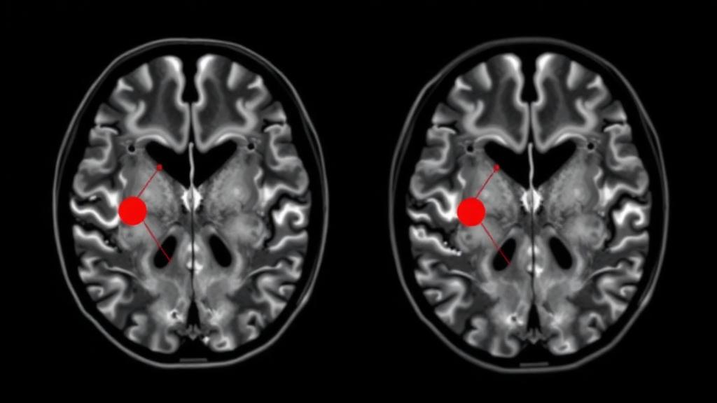
Diagnosing and grading a brain tumor from a magnetic resonance imaging scan is one of the most consequential tasks in clinical medicine, and it almost always depends on the trained eye of a specialist radiologist. That dependence on expert interpretation has long been a bottleneck: supervised artificial intelligence models that promise to automate tumor analysis need thousands of images annotated with detailed pathological grades, and such labels are scarce, expensive, and slow to produce. A new study published in Discover Artificial Intelligence by Nalineekumari Arasavali of Gayatri Vidya Parishad College of Engineering and colleagues proposes a way around the problem entirely, by asking a machine to organize brain tumor MRI scans into meaningful groups without ever being told what any of the images mean.
The researchers built an unsupervised framework that combines a deep autoencoder, a neural network trained to compress and then reconstruct its own input, with K-Means clustering, a classical algorithm that partitions data points by their distance to group centroids. The autoencoder first learns to squeeze each 224-by-224-pixel MRI image through a bottleneck of dense layers with 128 and then 64 ReLU-activated neurons, forcing the network to distill the scan into a compact latent vector. A mirrored decoder then attempts to rebuild the original image from that compressed code. By minimizing the reconstruction error between the original scan and its reconstruction, the network is pushed to preserve only the features that matter most: structural variations, intensity distributions, and texture-related abnormalities associated with tumor progression.
Once the autoencoder has been trained, the researchers discard the decoder and keep the encoder as a feature extractor. Every tumor image in their dataset, drawn from a publicly available Kaggle collection of 1,500 brain tumor scans, is passed through the encoder to produce a latent embedding. These embeddings, rather than the raw pixel values, are then fed into K-Means clustering with k-means++ centroid initialization, twenty initialization runs, and a fixed random seed for reproducibility. The result is a partition of the scans into three latent imaging-pattern groups, labeled Group 1, Group 2, and Group 3 purely for visualization purposes.
The authors are careful to stress that these three groups are not clinically validated tumor grades. Because the underlying dataset provides only tumor-versus-non-tumor labels rather than expert pathological grading, the clusters represent statistical similarities in imaging characteristics discovered by the algorithm, not medical severity categories. This distinction matters: the framework is positioned as an exploratory computational tool for pattern analysis, one that could support future research in settings where labeled data are limited, rather than as a diagnostic system ready for the clinic.
To judge whether the clustering was actually meaningful, the team benchmarked their approach against two popular hybrid alternatives: PCA plus K-Means, which uses classical linear dimensionality reduction, and VGG16 plus K-Means, which extracts features from a pre-trained convolutional neural network. Three standard internal validation metrics were computed for each method. The Silhouette Score measures how similar each point is to its own cluster compared with neighboring clusters, with higher values indicating better separation and compactness. The Calinski-Harabasz Index captures the ratio of between-cluster dispersion to within-cluster dispersion, again favoring higher values. The Davies-Bouldin Index, by contrast, rewards lower scores, since it quantifies average similarity between clusters based on inter- and intra-cluster distances.
The autoencoder-based method won on every metric. It achieved a Silhouette Score of 0.1601, a Calinski-Harabasz Index of 444.42, and a Davies-Bouldin Index of 1.939, the best results among all evaluated configurations. The VGG16- and PCA-based pipelines produced higher intra-cluster variance and weaker separation. The authors interpret these numbers as evidence that the deep learned features capture non-linear imaging characteristics that both linear PCA and transfer-learned CNN features miss. They also acknowledge that a Silhouette Score of roughly 0.16 indicates moderate separability, meaning some overlap persists between the discovered groups, a limitation they discuss openly.
Qualitative evidence came from t-distributed Stochastic Neighbor Embedding, or t-SNE, a technique that projects high-dimensional latent vectors into two dimensions for visualization. In the resulting plot, each point represents one MRI scan, colored by its assigned cluster. The three clusters appear as visibly distinct spatial groupings, supporting the idea that the autoencoder’s latent space organizes scans by imaging-pattern similarity. A bar chart of the cluster distributions further showed that the autoencoder-based method produced a more balanced spread of images across the three groups than either competing method, capturing more of the scans that the authors describe as representing more severe imaging patterns.
The study is candid about its limitations. The framework was validated on a single publicly available dataset, so cross-dataset and multi-institutional generalizability remain untested. Robustness against scanner variability, institutional differences, and varying noise conditions was not extensively evaluated. K-Means also assumes compact, roughly spherical clusters, which may not fully capture the complex, non-linear structures inherent in medical imaging data. And because the entire pipeline operates without clinical correlation or expert verification, direct medical interpretability remains limited. The clustering metrics measure computational separability, not diagnostic accuracy, and the authors explicitly warn that the identified groups should not be interpreted as clinically validated indicators of tumor severity or pathological grade.
The roadmap for future work is ambitious. The researchers plan to incorporate expert-annotated datasets containing pathological tumor grading, enabling them to test whether the latent imaging-pattern groups correspond to clinically validated grades when reviewed by medical professionals. They intend to explore probabilistic and density-based clustering methods, including Gaussian Mixture Models, DBSCAN, and spectral clustering, which may better model the messy distributions of real medical images. On the representation side, convolutional autoencoders, variational autoencoders, self-supervised learning, and contrastive representation learning are all on the table as ways to sharpen latent feature quality and cluster separability. The team also envisions integrating Explainable Artificial Intelligence techniques and quantitative tumor-region analysis to make the system’s groupings interpretable to clinicians.
What makes this work notable is its positioning within a broader shift in medical AI. As the authors note in their literature review, the field has been dominated by supervised deep learning, from CNN classifiers to VGG16-based hybrids and transformer models augmented with PCA, all of which demand extensive annotation. Unsupervised approaches like this one offer a complementary path: they can surface structure in unlabeled archives, flag candidate subgroups for expert review, and potentially reduce the annotation burden that has slowed clinical deployment of machine learning. By framing the output as exploratory imaging-pattern groups rather than diagnoses, the study models the kind of epistemic caution that unsupervised medical AI will need if it is ever to earn clinical trust. The work aligns with Sustainable Development Goal 3 on health and well-being and Goal 9 on industry, innovation, and infrastructure, and it suggests that even modest autoencoder architectures, when paired with careful validation, can extract genuine signal from the vast, unlabeled repositories of medical imaging data that hospitals accumulate every day.
Subject of Research: Unsupervised clustering of brain tumor MRI images using deep autoencoder embeddings and K-Means
Article Title: Unsupervised brain tumor MRI image grouping using deep autoencoder embeddings and K-means clustering
Article References: Arasavali, N., Gottapu, S., Sreelakshmi, K., Laveti, G., Nagothu, S. K., & Siddaraj, U. (2026). Unsupervised brain tumor MRI image grouping using deep autoencoder embeddings and K-means clustering. Discover Artificial Intelligence, 6(1), Article 1209. https://doi.org/10.1007/s44163-026-02241-9
Image Credits: AI Generated
DOI: 10.1007/s44163-026-02241-9
Keywords: brain tumor, MRI, autoencoder, K-Means clustering, unsupervised learning, medical imaging, t-SNE, Silhouette Score, PCA, VGG16, feature extraction, neuro-oncology
News Source: Nathaniel Bowman. (October 19, 2026). Deep Autoencoder Embeddings Sort Brain Tumor MRI Scans Without Labels. Scienmag.