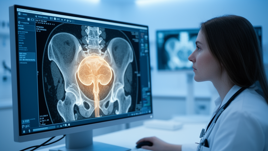
Bladder cancer has an awkward habit of coming back. Even after surgeons carefully scrape visible tumors from the bladder wall in a procedure known as transurethral resection, many patients find themselves back in the operating room within a couple of years. For the most common form of the disease, non-muscle-invasive bladder cancer, this pattern of recurrence is so frequent that doctors have long relied on statistical risk tables to decide who needs the most aggressive follow-up. Those tables, however, are built on a handful of clinical variables and leave enormous uncertainty around any individual patient. A new study published in BMC Medical Imaging suggests that the answer to this uncertainty may have been sitting inside the patient’s CT scan all along, waiting for the right algorithm to read it.
A team of researchers led by Zhan Jiang Yu and Zhe Hao Wu, working across three hospitals in Jiangxi Province, China, has built a deep learning model that ingests routine contrast-enhanced CT images and predicts whether a patient’s tumor will return within two years of surgery. The work is notable not just for its accuracy, which was substantial, but for its transparency. Using a mathematical technique called SHAP, the model can show clinicians exactly which features of the image and the patient’s clinical profile drove each prediction, addressing one of the most persistent objections to artificial intelligence in medicine: that neural networks are inscrutable black boxes whose advice cannot be trusted or audited.
The study enrolled 192 patients with non-muscle-invasive bladder cancer between June 2017 and May 2023. The largest hospital, the Second Affiliated Hospital of Nanchang University, contributed 139 patients who formed the training set, including 74 who experienced a recurrence. Two other centers, Jiangxi Cancer Hospital and a district hospital in Fuzhou, contributed 53 additional patients whose scans the model had never seen, providing an independent test set that included 24 recurrence events. This multicenter design matters. Models trained and tested on data from a single institution often fail when deployed elsewhere, because scanner settings, patient populations, and imaging protocols differ subtly from hospital to hospital. By validating on external data, the researchers gave their model a far more demanding examination.
Before any prediction could happen, the team had to confront a practical problem: CT images are not always sharp enough for fine-grained analysis. Their solution was to apply super-resolution image reconstruction powered by a generative adversarial network, a class of artificial intelligence in which two neural networks compete against each other, one generating enhanced images and the other trying to detect the fakes, until the generator produces images detailed enough to fool its opponent. Applied to the contrast-enhanced CT scans, this technique recovered fine tissue textures that standard reconstruction can blur away, giving the downstream models richer information to work with. It is a clever piece of engineering that turns an ordinary diagnostic scan into a high-fidelity substrate for machine learning.
From these enhanced images, the researchers extracted two complementary kinds of information. The first was radiomics: hundreds of quantitative measurements describing tumor texture, shape, and intensity patterns, such as features derived from gray-level co-occurrence matrices and neighboring gray-tone difference matrices, which capture how pixel intensities repeat and cluster within the tumor. These handcrafted features encode characteristics that the human eye perceives only vaguely as a tumor looking heterogeneous or mottled. The second source was deep learning features, learned automatically by a convolutional neural network that processed the images directly and distilled them into abstract numerical representations. Finally, the team compiled clinical variables, including tumor number and T stage, the standard measure of how deeply the tumor has invaded the bladder wall.
The centerpiece of the study is what the authors call Fusion Model C, which combines all three information streams. Trained exclusively on the first center’s data, it achieved an area under the receiver operating characteristic curve of 0.917 in the training set, with a 95 percent confidence interval of 0.874 to 0.960, and 0.865 on the unseen external test set, with a confidence interval of 0.756 to 0.974. For context, an AUC of 0.5 reflects random guessing, while 1.0 reflects perfect discrimination, so a value of 0.865 on external data represents strong, clinically meaningful performance. The fused model clearly outperformed a radiomics-only model, which managed an AUC of 0.754 on the test set, demonstrating that the deep learning features and clinical variables carried predictive power that texture statistics alone could not supply.
Accuracy alone, however, was not the study’s only ambition. The researchers interrogated their model with Shapley additive explanations, a technique borrowed from cooperative game theory that assigns each input feature a precise contribution to every individual prediction. The analysis revealed that tumor number, T stage, and a single deep learning feature were the three highest-ranked predictors overall. In other words, the model had independently learned something the clinical literature already suspected, that patients with multiple tumors and higher T stages recur more often, while also discovering predictive image patterns that no traditional risk table captures. Calibration curves, Brier scores, and decision-curve analysis further supported the model’s reliability, with the decision curves in particular suggesting that using the model to guide surveillance decisions would yield more net benefit than treating all patients identically or treating none.
The clinical implications are significant because surveillance for non-muscle-invasive bladder cancer is burdensome and expensive. Current practice relies on risk stratification systems from the European Organization for Research and Treatment of Cancer and the Spanish Urological Oncology Treatment Group, which combine factors such as tumor count, size, stage, grade, and prior recurrence rate to sort patients into risk categories. These categories determine how often patients undergo cystoscopy, an uncomfortable camera examination of the bladder, and whether they receive intravesical therapies such as Bacillus Calmette-Guérin instillations. A model that reads a preoperative CT scan, which many patients already undergo, could refine this stratification before any treatment decision is made, potentially sparing low-risk patients some procedures while flagging high-risk patients for intensified follow-up.
The study’s authors are careful about scope, and rightly so. The work is retrospective, meaning the researchers analyzed scans and outcomes that had already been collected rather than following new patients forward in time. The test set, while external, contained only 53 patients and 24 recurrence events, which widens the confidence intervals and means the reported performance carries real statistical uncertainty. The model was also developed and validated in a specific Chinese patient population, and its behavior in other populations, with different scanners and ethnic profiles, remains to be demonstrated. Prospective validation, in which the model’s predictions are locked in before outcomes are known, would be the natural next step before any deployment in clinical practice.
Even with those caveats, the study offers a compelling glimpse of where cancer surveillance is heading. The combination of generative adversarial super-resolution, radiomics, deep learning, and explainable artificial intelligence transforms a routine preoperative scan into a personalized prognosis engine, one that can justify its conclusions to the physicians who must act on them. The research was supported by the Jiangxi Provincial Health Commission, the Jiangxi Provincial Academic and Technical Leader Training Program, and the National Natural Science Foundation of China, and the authors report no competing interests. If future prospective studies confirm these results, the two-year question that haunts every bladder cancer patient leaving the operating room may one day be answered, with quantified confidence, before the patient even wakes up.
Subject of Research: An explainable deep learning model using contrast-enhanced CT to predict two-year recurrence of non-muscle-invasive bladder cancer
Article Title: An interpretable deep learning model based on enhanced CT predicts the recurrence of non-muscle-invasive bladder cancer within 2 years
Article References: Yu, Z. J., Wu, Z. H., Huang, J. B., Zeng, Z. F., Chao, H. C., & Zeng, T. (2026). An interpretable deep learning model based on enhanced CT predicts the recurrence of non-muscle-invasive bladder cancer within 2 years. BMC Medical Imaging. https://doi.org/10.1186/s12880-026-02738-w
Image Credits: AI Generated
DOI: 10.1186/s12880-026-02738-w
Keywords: bladder cancer, NMIBC, deep learning, radiomics, contrast-enhanced CT, generative adversarial network, super-resolution, SHAP, recurrence prediction, machine learning, medical imaging, predictive medicine
News Source: Nathaniel Bowman. (October 18, 2026). AI Reads CT Scans to Predict Bladder Cancer Recurrence Before It Happens. Scienmag.