For nearly a thousand years, practitioners of traditional Chinese medicine have prized Chansu, a dried venom secreted by the ears and skin glands of two Asian toad species, as a remedy for swelling, pain, heart failure, and cancer. Now a research team at Shandong Medical and Pharmaceutical University and Qingdao Central Hospital has traced one of the venom’s most potent anticancer ingredients to a surprising molecular destination: the peroxisome, a tiny organelle that most cell biologists once dismissed as little more than a cellular janitor. In a study published in Frontiers in Pharmacology, the team shows that cinobufagin, a bufadienolide steroid from Chansu, collapses the biogenesis machinery of peroxisomes in liver cancer cells, starving the cells of the lipid metabolic flexibility they need to grow.
The stakes are considerable. Primary liver cancer is the sixth most commonly diagnosed cancer worldwide and the third leading cause of cancer death, with roughly 906,000 new cases and 830,000 deaths recorded in 2020 according to the Globocan report. Hepatocellular carcinoma, the dominant subtype accounting for 75 to 85 percent of cases, is notoriously difficult to treat. Seventy to eighty percent of patients are diagnosed at middle or advanced stages, when surgical resection is no longer an option, and chemotherapy drugs tend to cause serious side effects while delivering unsatisfactory efficacy. Current first-line regimens built around sorafenib tosylate and durvalumab-based immunotherapy help some patients, but the search for new, low-toxicity anticancer drugs remains urgent.
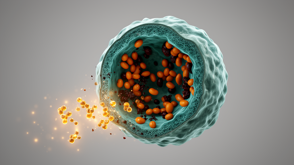
Peroxisomes have spent decades in the shadow of mitochondria. First discovered in 1954 and formally named as an organelle in 1966, they were long thought of as the cellular hoi polloi, scavenging reactive oxygen debris discarded by more glamorous organelles. But their function extends far beyond decomposing hydrogen peroxide. Peroxisomes house the catabolism of very long chain fatty acids, the synthesis of plasmalogens and ether phospholipids, and a battery of matrix enzymes, of which catalase is the hallmark, making up about 40 percent of total peroxisomal protein. Their biogenesis depends on a family of proteins called peroxins, encoded by more than 30 PEX genes, including PEX5, the cytosolic receptor that ferries matrix enzymes to the peroxisome membrane, and PEX10, part of the membrane-bound ubiquitin ligase complex that recycles PEX5 after each delivery round.
Recent work has begun to reposition peroxisomes as active players in cancer. Enzymes involved in peroxisomal lipid processing are elevated in numerous tumor types, and studies in mice have shown that tumors shrink when peroxisomal lipid processing is inhibited or genes tied to peroxisomal degradation are silenced. High expression of PEX5 has been linked to poor prognosis in hepatocellular carcinoma patients, where ectopic PEX5 increases proliferation, migration, and invasiveness. The survival of liver cancer cells, in short, may depend on peroxins. That dependency makes the peroxisome biogenesis pathway an attractive therapeutic target, and it is precisely where cinobufagin strikes.
The team’s earlier transcriptome sequencing of cinobufagin-treated HepG2 cells, a widely used human hepatoma line, hinted at the mechanism. KEGG enrichment analysis of the differentially expressed genes revealed that peroxisome intervention was partially responsible for the drug’s antitumor effects. In the new study, the researchers confirmed the hit with Western blotting and immunofluorescence. Cinobufagin significantly reduced the expression of catalase and ABCD3, a 70-kilodalton peroxisomal membrane protein used as a standard marker of peroxisome number, in both HepG2 and SK-HEP-1 cells. Confocal imaging showed a marked drop in average ABCD3 fluorescence intensity, indicating fewer peroxisomes per cell. The drug also downregulated PEX5, PEX6, and PEX10 at the protein level, with transcriptomic data showing log2 fold changes of minus 1.83, minus 3.36, and minus 2.75 respectively.
To test whether those peroxins actually drive the drug’s growth-inhibitory effect, the team silenced PEX5, PEX6, or PEX10 individually with small interfering RNA. Knocking down PEX5 or PEX10 significantly suppressed proliferation and colony formation in both cell lines, and depleting either peroxin enhanced cinobufagin’s blocking effect on cell growth. PEX6 knockdown, by contrast, had no measurable effect on proliferation, a discrepancy the authors attribute to functional specificity among PEX family members or cell type specificity. The biological significance of cinobufagin’s inhibition of PEX6, they suggest, may not lie in suppressing tumor cell proliferation directly.
The peroxisome connection then led the team to lipid metabolism, the organelle’s other major portfolio. Cinobufagin restrained the expression of three lipid metabolic enzymes: fatty acid synthase, or FASN, and ATP-citrate lyase, or ACLY, both overexpressed in most cancer cells and positively correlated with malignant proliferation and poor prognosis, and ECH1, a key enzyme in fatty acid beta-oxidation present in both peroxisomes and mitochondria. Mechanistic experiments showed that PEX5 knockdown inhibited FASN and ACLY expression, while PEX10 knockdown suppressed FASN alone, implying that cinobufagin restrains lipid metabolism partially through suppression of PEX5 and PEX10. An oral FASN inhibitor, TVB2640, is already in clinical trials for liver cancer, underscoring the therapeutic relevance of this axis.
Untargeted metabolomics of cinobufagin-treated HepG2 cells then mapped the downstream metabolic wreckage. Of the lipid metabolites detected, glycerophospholipids accounted for 44 percent and fatty acyls for 38 percent. Cinobufagin reduced the levels of 12 fatty acyl species, including elaidic acid, mevalonic acid, and several acylcarnitines, and suppressed dozens of glycerophospholipids, including 13 lysophosphatidyl ethanolamines, 14 lysophosphatidyl cholines, and 4 lysophosphatidyl serines. Those lysolipids are not passive bystanders: LPC has been reported to stimulate tumor cell migration through G-protein coupled receptors, LPE promotes migration and proliferation via MAPK signaling, and LPS drives chemotactic migration through GPR34 and the PI3K/AKT pathway. Their coordinated collapse suggests that cinobufagin interferes with the lipid metabolic reprogramming that fast-growing tumors rely on.
In vivo, the team inoculated Kunming mice with murine H22 hepatoma cells and treated them intraperitoneally with cinobufagin at 1 or 2 milligrams per kilogram per day for 13 days. Tumor growth was significantly restrained, with no significant weight loss, no histological signs of toxicity in the heart, liver, spleen, lung, or kidney, and no significant changes in serum ALT or AST, indicating preserved liver function. Hematoxylin and eosin staining of tumor sections showed reduced cell density, nuclear contraction and rupture, and increased connective tissue proliferation in the cinobufagin groups, with necrosis increasing at higher doses. Serum metabolomics revealed that 24 lipid and lipid-like molecules changed significantly, including 14 fatty acyls, six glycerophospholipids, one sphingolipid, two sterols, and one prenol lipid, with phosphocholine, a glycerophospholipid metabolic intermediate, markedly reduced.
Some of the serum shifts carry clinical echoes. Cinobufagin lowered phosphatidyl ethanolamine 18:1_18:1, a species whose serum levels are elevated in hepatocellular carcinoma patients compared with those with cirrhosis, and reduced sphingomyelin SM 8:1; 2O/26:0, a class of lipids increasingly recognized as diagnostic markers for HCC. It also raised levels of 25-hydroxycholecalciferol, the liver-derived prohormone whose deficiency is associated with advanced HCC stage and poor prognosis, and vitamin A, whose low levels have been linked to hepatitis C, cirrhosis, and HCC risk. The authors conclude that cinobufagin is a promising natural drug candidate for liver cancer treatment and that PEX proteins may be its molecular targets. The work, they write, lays a theoretical foundation for the clinical application of cinobufagin and Chansu, and promotes the development of new drugs from traditional Chinese medicine.
Subject of Research: Cell Biology
Article Title: Cinobufagin induces peroxisome dysfunction through suppression of the peroxisomal biogenesis factor proteins and interferes with lipid metabolism to affect the growth of liver cancer cells
Article References: Yang, J., Shao, W., Yu, C., Wu, Q., Xu, R., Rong, X., & Yang, A. (2026). Cinobufagin induces peroxisome dysfunction through suppression of the peroxisomal biogenesis factor proteins and interferes with lipid metabolism to affect the growth of liver cancer cells. Frontiers in Pharmacology, 17, Article 1860569. https://doi.org/10.3389/fphar.2026.1860569
Image Credits: AI Generated
DOI: 10.3389/fphar.2026.1860569
Keywords: cinobufagin, peroxisome, liver cancer, lipid metabolism, PEX5, PEX10, Chansu, metabolomics, hepatocellular carcinoma, natural products, induces, dysfunction
News Source: Nathaniel Bowman. (October 17, 2026). Toad Venom Compound Cinobufagin Disables Liver Cancer Cells by Shutting Down Their Peroxisomes. Scienmag.