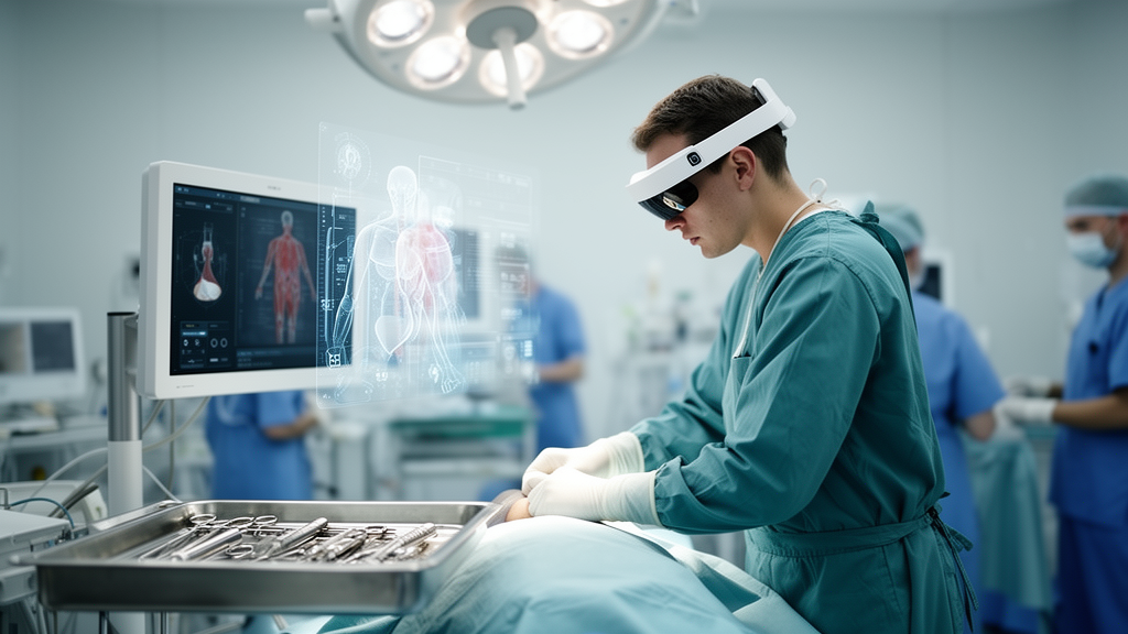
When Apple released the Vision Pro in early 2024, it marketed the device as a consumer product for entertainment and productivity. Three years later, a team of Chinese researchers has compiled the most comprehensive picture yet of how surgeons, radiologists, and medical educators have quietly put the headset to work in hospitals around the world. Their scoping review, published in Frontiers in Medicine, examined 69 studies and found that the device has been tested in everything from laparoscopic colorectal cancer surgery to cataract operations, liver tumor ablation, and anatomy teaching. But the review also delivers a sobering caveat: nearly all of the evidence comes from feasibility studies, case reports, and opinion pieces, and only two confirmatory trials have been conducted to date.
The Vision Pro is Apple’s first head-mounted spatial computing device, officially announced on June 6, 2023, and launched in the United States on February 2, 2024. It weighs between 600 and 650 grams and is powered by two chips: the M2, which handles general computing, and the R1, which processes sensor data with a cited photon-to-photon latency of under 12 milliseconds. The exterior features a glass panel and a 4K Micro-OLED display capable of brightness up to 5,000 nits and a resolution of 23 million pixels. An integrated 3D camera captures depth photographs and 3D videos, while 12 external cameras, a high-precision eye-tracking system, and a multi-sensor array enable ultra-high-resolution, low-latency passthrough of the real world and spatial positioning of virtual content. The device’s defining feature is its comprehensive integration of augmented and virtual reality, allowing real-time multi-screen data overlay and interaction with 3D models. In clinical settings, this means surgeons can seamlessly integrate preoperative imaging, intraoperative real-time data, and electronic medical records into a single field of view.
The review’s authors, led by Yiwei Hu of the First Affiliated Hospital with Nanjing Medical University and Chuxu Wang of Huai’an Second People’s Hospital, conducted their search across PubMed, Web of Science, Scopus, and the Chinese Medical Association Journal Database, covering publications from January 1, 2023, to September 9, 2026. After removing duplicates and screening 251 records, they included 69 articles in the final analysis. To assess the maturity of the evidence, they applied the IDEAL-D framework, a standardized pipeline for medical device innovation. They classified 10 studies as Stage 0 preclinical exploratory work using phantoms or ex vivo tissues, 23 as Stage 1 or 2a first-in-human or exploratory studies, and just 2 as Stage 2b confirmatory studies with controlled designs. The remaining 34 articles, comprising medical education studies without original patient data and opinion pieces, fell outside the framework. This distribution means the overall body of evidence remains dominated by early-stage technical demonstrations rather than rigorous clinical trials.
In the operating room, the Vision Pro has been most extensively tested as a visualization platform for minimally invasive surgery. Shen and colleagues described a connection method in which a capture card was linked to the video output of a laparoscope and then to a MacBook via USB-C, enabling real-time transmission of laparoscopic video to the headset. Under default settings, the latency was approximately 120 milliseconds, which could be improved to under 100 milliseconds with resolution adjustment. The same team later optimized the architecture using a Blackmagic Design capture device connected to a Mac Studio via Thunderbolt 3, with a 10 GbE Ethernet link to a high-end router, controlling transmission latency to within 60 milliseconds. Surgeons could then observe a highly synchronized real-time 3D surgical field through the headset. In a series of 22 laparoscopic colorectal cancer surgeries, the mean operative time was 189 minutes, no intraoperative complications occurred, and System Usability Scale scores ranged from 90 to 100 across varying surgical difficulty.
Broader surgical series have reinforced these findings. Broderick’s team at the University of California, San Diego, employed the Vision Pro as the primary monitor in 41 minimally invasive general surgeries, allowing clinicians to view up to three virtual displays simultaneously and adjust their position and size according to individual preference. The mean NASA Task Load Index score for the entire cohort was 22.3 out of 100, indicating low subjective cognitive load, with no significant difference between attending surgeons and trainees. No intraoperative complications occurred, and the 30-day morbidity and mortality rates were both zero. In spinal endoscopic surgery, Park and colleagues combined augmented reality with lumbar stenosis treatment, displaying real-time endoscopic video alongside electronic medical records and preoperative MRI in the center of the field of view. This reduced cervical strain and improved decision-making safety. A larger prospective study in 40 patients undergoing biportal endoscopic lumbar surgery showed a mean Surgery Task Load Index score of 22.24, with low complication rates and early outcomes comparable to conventional surgery.
Ophthalmic surgery has emerged as another promising domain. Orione and colleagues were the first to apply the Vision Pro in corrective surgery for eyelid malposition, with ten ophthalmologists completing procedures while wearing the headset throughout. Surgeons overlaid preoperative patient images in real time on one side of the surgical field while simultaneously displaying a surgical step checklist, the local anesthetic effect curve, and a timer on the other. All System Usability Scale scores exceeded 85, and no complications occurred. Mastropasqua and colleagues extended the application to more delicate intraocular procedures, including phacoemulsification with intraocular lens implantation, silicone oil removal, and vitrectomy with epiretinal membrane peeling. Using the device’s multimodal image fusion function, surgeons could retrieve preoperative fundus autofluorescence, optical coherence tomography, and fundus photography images at any time during surgery. All procedures were completed successfully with a zero percent intraoperative and short-term postoperative complication rate. The same study explored a remote surgical guidance model, connecting five trainee physicians at varying distances via FaceTime to share the surgical field of view in real time, with no significant signal latency observed.
In diagnostic radiology, Murphy and colleagues conducted a pilot study suggesting that the Vision Pro’s primary application scenarios include the interpretation of abdominal and pelvic CT images and the 3D reconstruction of complex anatomical structures. For CT-based diagnosis of diverticulitis, the area under the curve was 0.93, which was not statistically significantly different from that of desktop monitors at 0.94. The device demonstrated significant advantages in immersion and intuitiveness when used for 3D reconstruction, with a mean preference score of 3.3 on a 5-point scale. However, the median diagnostic time per case was 57 seconds, considerably longer than the 31 seconds for desktop monitors, and the device lacked calibration for the DICOM Grayscale Standard Display Function. As a result, radiologists still preferred conventional monitors for basic image viewing scenarios, though the Vision Pro showed clear advantages in the 3D visualization of complex anatomical relationships.
In medical education, the Vision Pro has been applied across anatomy teaching, clinical skills training, specialized procedural simulation, and distance learning. The device can overlay digital anatomical models onto real-world scenarios, circumventing issues of cadaver shortage, ethical controversy, and high cost while enhancing learners’ comprehension of intricate anatomical relationships through interactive manipulation. Applications such as INSIGHT HEART deliver dynamic, stereoscopic, and dissectible cardiac models that vividly illustrate the entire cardiac pumping cycle. In surgical training, Shen and colleagues reported that 50 trainees improved operative skills by 15 percent and exam scores by 9 percent, with 88 percent reporting enhanced professional identity. Stauffer and colleagues developed an augmented reality-enhanced physical simulator for femoral nailing using the Vision Pro as the display platform, in which 23 participants completed three unguided simulation runs followed by a retention session. Task completion time decreased by 47.1 percent for insertion and 57.4 percent for alignment across runs, while the number of simulated fluoroscopic images declined by 45.6 percent and 49.2 percent respectively. In distance education, Goh and colleagues utilized an indoor 5G small-cell architecture to transmit the high-definition 3D view from a da Vinci surgical robot during transoral robotic surgery to the Vision Pro headsets of remote trainees, with a mean latency of 22 milliseconds. Twenty trainees from seven countries obtained a stereoscopic, immersive viewing experience approximating the first-person perspective of the operator.
Despite these encouraging signals, the review identifies substantial limitations that temper any suggestion of imminent clinical transformation. The device weighs between 600 and 650 grams, and clinical studies suggest that prolonged wearing often causes cervical fatigue in surgeons. Minor alterations in headset position significantly impair the precision of eye-tracking, leading to discrepancies between virtual content and actual anatomical structures. Its battery life of roughly two hours is insufficient for lengthy surgical procedures, and the 2025 hardware update, while enhancing chip performance and extending battery duration, added 150 grams of weight. The device has not obtained marketing approval from major regulatory bodies such as the FDA or NMPA as a diagnostic or surgical navigation device, and it lacks DICOM calibration for primary diagnostic interpretation. Compatibility with existing hospital information systems, electronic medical records, and picture archiving systems remains suboptimal. The device is priced at approximately 3,500 dollars, with hidden costs including specialized software development, system maintenance, personnel training, and workflow redesign further increasing the economic burden on healthcare institutions. No mainstream reimbursement system currently covers Vision Pro-assisted surgery.
Perhaps the most instructive contradiction arises from two phantom studies of percutaneous puncture navigation. In a direct comparative benchmark of HoloLens 2, Magic Leap 2, and the Vision Pro for needle insertion, the Vision Pro achieved the smallest distance error during successful punctures at 6.34 millimeters but an overall success rate of only 52.46 percent, with lateral errors in failed attempts reaching 19.39 millimeters. User feedback cited unstable marker tracking, excessive weight, and incompatibility with prescription glasses as major obstacles. By contrast, Lan and colleagues, using a Vision Pro-based real-time image fusion navigation system for laparoscopic microwave ablation of hepatic hemangiomas, reported a puncture success rate of 90 percent under Vision Pro guidance compared with 42.5 percent using conventional methods. The review’s authors attribute this discrepancy not to a failure of the hardware itself but to fundamental differences in the navigation paradigm: dynamic instrument tracking is highly sensitive to headset displacement and tracking drift, while static image fusion places lower demands on spatial registration stability. This suggests a critical hierarchy of suitability, with the device currently performing well for static image reference applications and moderately for 3D overlay guidance, but remaining immature for dynamic surgical navigation with instrument tracking. The authors conclude that widespread adoption should not occur until Stage 2b trials confirm non-inferiority or superiority in patient-relevant outcomes, and they call for multicenter randomized controlled trials, standardized operating procedures, and long-term registry studies to transition the Vision Pro from technical demonstration to evidence-based clinical practice.
Subject of Research: Health
Article Title: Early applications of apple vision pro in clinical medicine and medical education: a scoping review
Article References: Hu, Y., Wang, Y., Zuo, X., Hu, H., Min, B., Wang, C., Tang, Z., Xiao, N., Huang, C., & Wang, C. (2026). Early applications of apple vision pro in clinical medicine and medical education: a scoping review. Frontiers in Medicine, 13, Article 1909038. https://doi.org/10.3389/fmed.2026.1909038
Image Credits: AI Generated
DOI: 10.3389/fmed.2026.1909038
Keywords: Apple Vision Pro, mixed reality, surgery, medical education, spatial computing, clinical medicine, Frontiers in Medicine, Early, applications, apple, vision, clinical
News Source: Ophelia Keating. (October 17, 2026). Apple Vision Pro Shows Early Promise in Surgery and Medical Education, but Evidence Remains Thin. Scienmag.