


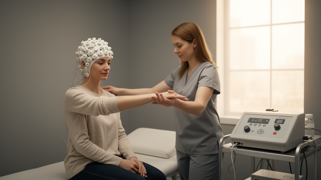
When a 39-year-old agricultural worker found his body refusing to obey him, doctors faced one of the most puzzling conditions in modern neurology. His arms trembled violently whenever he tried to raise them, his legs shook when he walked, and he struggled with unexplained fevers, migraines, and blood pressure spikes. Now, in a remarkable case study published in Frontiers in Psychiatry, researchers report how they used electroencephalography (EEG) to guide low-frequency repetitive transcranial magnetic stimulation (rTMS) targeting the supplementary motor area (SMA), followed by staged rehabilitation, to help this patient regain control of his movements.
Functional movement disorder (FMD) is a subtype of functional neurological disorder characterized by abnormal movements, including functional tremors, dystonia, gait disturbances, and parkinsonism, that are not explained by structural damage or another movement disorder. Historically dismissed as purely psychological, FMD is increasingly recognized as a genuine disorder of brain network function. Recent neuroimaging studies have revealed altered connectivity within motor networks, particularly involving the SMA and primary motor cortex, suggesting that the condition reflects measurable changes in how brain regions communicate rather than imagined symptoms.
The patient’s story began when he developed progressive tremors and co-contraction of his arms and legs that interfered with daily activities. Despite extensive testing, including brain MRI, blood work, and nerve conduction studies, no structural cause could be identified. He also experienced new-onset migraines, recurrent fevers, and hypertension, complicating the diagnostic picture. After years of unsuccessful treatments, he was referred to the researchers’ hospital for evaluation of rTMS therapy.
Before stimulation, the team recorded resting-state EEG under eyes-closed conditions and analyzed functional connectivity between electrode sites. They found relatively prominent theta activity at the Cz electrode, overlying the medial frontal cortex, together with altered alpha synchronization patterns across frontal and parietal regions. The researchers interpreted these findings as evidence of hyperexcitability within medial motor regions, consistent with the known role of the SMA in generating functional tremors. Based on this EEG profile and the established literature, they selected the SMA as the stimulation target.
The team applied low-frequency rTMS, delivering 1,200 pulses per session at 90% of the resting motor threshold, over the SMA. Low-frequency stimulation is thought to suppress cortical excitability, potentially normalizing the hyperactive medial motor circuits implicated in functional tremors. The patient underwent ten sessions over two weeks, followed by staged rehabilitation that progressed from relaxation and gentle passive movements to active task-oriented exercises incorporating video feedback of his own movements.
The results were striking. By the end of treatment, the patient’s tremors during bilateral arm elevation had diminished noticeably, and he could perform daily activities with greater ease. Standardized assessments confirmed improvements in the FMD rating scale and activities of daily living. Perhaps most remarkably, follow-up EEG revealed normalization of the previously altered connectivity patterns, with reduced relative prominence of the Cz theta activity and more balanced alpha synchronization across frontal and parietal sites. The brain’s communication patterns appeared to shift toward normal as movements improved.
The researchers emphasize that this case illustrates the potential of EEG-guided neuromodulation for FMD. Rather than applying stimulation blindly, the team used the patient’s own brain activity to identify hyperexcitable regions and select an individualized target. This approach aligns with the emerging concept of brain-state-dependent stimulation, in which the patient’s neural profile determines the intervention. The staged rehabilitation that followed may have consolidated the gains by retraining motor circuits through repeated, feedback-enhanced practice.
However, the researchers caution that this is a single case study and that the tremor recurrence at follow-up highlights the need for longer-term strategies. The patient’s tremors returned within weeks of discharge, suggesting that the neural changes induced by stimulation may require maintenance sessions or ongoing rehabilitation to sustain. The researchers also note that the autonomic symptoms, including fevers and blood pressure fluctuations, remained unexplained and may reflect broader network dysfunction beyond the motor system.
Despite these limitations, the case offers a compelling glimpse of the future of FMD treatment. As neuroimaging and EEG analysis become more sophisticated, clinicians may increasingly use patients’ own brain activity to guide individualized neuromodulation, moving beyond the one-size-fits-all approaches that have dominated the field. For a condition long dismissed as untreatable or imagined, the ability to visualize and reshape brain network function represents a genuine breakthrough, offering hope to patients whose bodies have betrayed them for years.
The epidemiological profile of FMD helps place this case in context. With an annual incidence of roughly 4 to 5 per 100,000 people and a prevalence near 50 per 100,000, the condition is far from rare, and it is frequently encountered in general neurology clinics and pediatric departments. Functional tremor, the subtype seen in this patient, is the most common presentation. Although approximately 70 percent of affected individuals are women, with a mean age at onset of around 40 years, the proportion of male patients rises with age, which is relevant when considering cases like this one involving a man in his late thirties.
The precipitating event described here also fits a well-recognized pattern. Patients with FMD often report psychological stressors or physical trauma, such as motor vehicle accidents, in the period preceding symptom onset. In this instance, a load weighing more than 100 kilograms fell toward the patient at work, twisting his body forcibly to the left and triggering severe neck and back pain. Structural workup, including lumbar MRI showing a sacral bone contusion and disc herniation, could not account for the generalized movement disorder that emerged, and the subsequent development of tremors, abnormal sensations, fever, and hypertension one month after injury illustrates how functional symptoms can layer onto genuine tissue injury, complicating diagnosis and delaying recognition.
The autonomic dimension of this case deserves particular attention. Beyond motor symptoms, FMD has been associated with alterations in autonomic and endocrine function, and recent studies have reported diminished vagal tone in these patients, suggesting altered autonomic regulation and reduced stress resilience. The patient’s unexplained hypertension, recurrent fevers of 37 to 38 degrees Celsius, and new-onset migraine prompted an unusually thorough differential workup, including serum catecholamine measurements to exclude pheochromocytoma, autoimmune screening, head-up tilt testing, and sympathetic skin response testing. Notably, heart rate variability was not assessed, which the authors acknowledge as a limitation given that reduced vagal tone has been documented in FMD populations and might have provided a physiological correlate of the autonomic instability observed.
The neuroimaging literature offers a framework for understanding why the supplementary motor area was chosen as the stimulation target. In patients with functional tremor, prior multimodal imaging work has described increased activity within a motor-limbic network involving the anterior cingulate cortex and paracingulate cortex, alongside reduced activation of the right temporoparietal junction. This reduced temporoparietal activation is thought to contribute to an impaired sense of agency, the subjective experience that movements occur outside one’s voluntary control, which is a hallmark complaint in FMD. Increased resting-state regional cerebral blood flow has also been reported in the left inferior frontal gyrus and insular cortex. These findings collectively portray FMD as a disorder of distributed network dynamics rather than a focal lesion, providing a rationale for using connectivity measures to guide intervention.
The EEG methodology used in the case reflects this network perspective. Resting-state recordings were acquired with a standard clinical amplifier using the international 10-20 electrode system, with averaged auricular references, under eyes-closed conditions. After excluding artifact-contaminated segments, the team analyzed a continuous five-minute segment using power spectral density estimation via Welch’s method and the weighted phase lag index, a connectivity measure that is relatively robust to volume conduction artifacts. Analyses were performed with established open-source Python libraries, and spectral estimates were normalized across the frequency range to allow comparison between electrodes. This combination of spectral and connectivity metrics allowed the team to characterize both local oscillatory abnormalities and the coordination between distant regions, which is precisely the kind of information needed to select a neuromodulation target in a network disorder.
The rehabilitation component of the intervention is also grounded in established principles. Staged rehabilitation that progresses from relaxation and passive movement toward active, task-oriented exercise with visual feedback exploits the same neuroplastic mechanisms that underlie motor learning in other neurological conditions. Visual feedback of one’s own movement is particularly relevant in FMD, where restoring the sense of agency over voluntary action is considered a key therapeutic goal. The combination of neuromodulation to reduce abnormal excitability and structured practice to reinforce adaptive motor patterns represents a theoretically coherent pairing, even though the relative contributions of each component cannot be disentangled in a single case.
The recurrence of tremors after discharge underscores a broader challenge in FMD management. Symptom relapse following short courses of intervention is common across treatment modalities, and the finding that gains eroded within weeks suggests that network changes induced by stimulation may be functionally reversible rather than structurally consolidated. Maintenance stimulation schedules, extended rehabilitation, or periodic reassessment with EEG to retune targets may be needed to sustain benefit. The authors’ decision to keep all regularly scheduled medications, including those for dilated cardiomyopathy and migraine, at unchanged doses throughout the treatment period strengthens the attribution of motor improvements to the neuromodulation and rehabilitation program rather than to pharmacological changes.
Ultimately, this case adds to a growing body of literature suggesting that FMD, long dismissed as untreatable or imagined, can be approached with the same objective, mechanism-targeted tools applied to other neurological disorders. The convergence of EEG-based network analysis, rational target selection, and feedback-enhanced rehabilitation offers a template for future controlled studies, which will be essential to determine whether such individualized protocols can deliver durable benefit across the broader FMD population.
Subject of Research: Case Report: Electroencephalography-informed low-frequency repetitive transcranial magnetic stimulation targeting the supplementary motor area in severe functional movement disorder
Article Title: Case Report: Electroencephalography-informed low-frequency repetitive transcranial magnetic stimulation targeting the supplementary motor area in severe functional movement disorder
Article References: Takahashi, C., Asahi, T., Kanda, M., Ohta, Y., & Matsushita, I. (2026). Case Report: Electroencephalography-informed low-frequency repetitive transcranial magnetic stimulation targeting the supplementary motor area in severe functional movement disorder. Frontiers in Psychiatry, 17, Article 1959751. https://doi.org/10.3389/fpsyt.2026.1959751
Image Credits: AI Generated
DOI: 10.3389/fpsyt.2026.1959751
Keywords: functional movement disorder, transcranial magnetic stimulation, EEG, supplementary motor area, neuromodulation, brain connectivity, rehabilitation, case study, Case, Report, Electroencephalography-informed, low-frequency
News Source: Cassandra Pierce. (October 17, 2026). EEG-Guided Brain Stimulation Rebuilds Movement in Severe Functional Movement Disorder Case. Scienmag.


