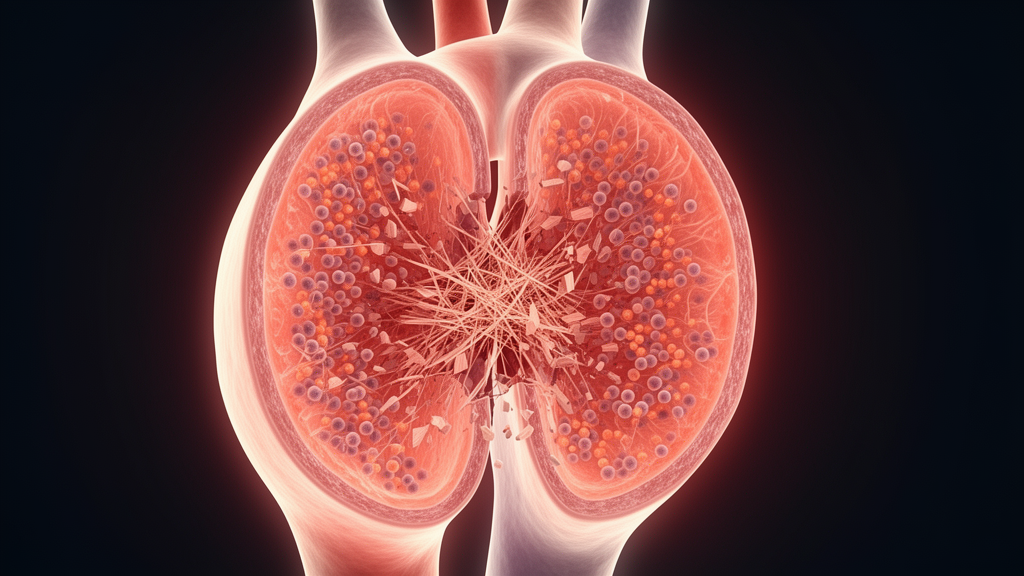
For decades, the thickening and stiffening of the aortic valve that precedes clinically significant stenosis was dismissed as an inevitable consequence of aging, a passive wearing-down of tissue that required no more attention than gray hair or wrinkled skin. A new study from the National Institute of Cardiology Ignacio Chávez in Mexico City challenges that view at the molecular level. Reporting in the British Journal of Biomedical Science, a research team led by Elizabeth Chan-Delgado and Luis M. Amezcua-Guerra shows that aortic valve sclerosis, the early and often asymptomatic stage of calcific aortic valve disease, carries a distinctive and active biological signature: a surge in the matrix-degrading enzyme MMP-9, a drop in its natural inhibitor TIMP-1, and elevated levels of the inflammatory cytokine tumor necrosis factor. The findings suggest that the early phase of valve disease is not a quiet precursor but a biologically turbulent process, one that may be amenable to intervention before irreversible calcification sets in.
Calcific aortic valve disease exists on a continuum. It begins with aortic valve sclerosis, in which the valve leaflets thicken and develop focal areas of increased echogenicity on ultrasound, yet blood flow remains unobstructed and echocardiographic parameters stay within normal limits. Over years, this subclinical state can progress to aortic stenosis, the advanced hemodynamically significant phase in which the valve area shrinks, transvalvular pressure gradients climb, and the heart must strain to push blood through a narrowed orifice. The epidemiology is sobering. The prevalence of aortic stenosis rises from roughly 0.2 percent among people aged 50 to 59 to nearly 10 percent among those aged 80 to 89, while aortic valve sclerosis affects an estimated 25 to 40 percent of individuals between 65 and 75. Because sclerosis is usually silent, most patients are identified only once significant stenosis has developed, at which point surgical or interventional valve replacement remains the sole effective therapy. Untreated severe symptomatic stenosis carries a high mortality, making the search for early molecular markers and therapeutic targets a clinical priority.
The conceptual framework behind the new work draws heavily on atherosclerosis. Calcific aortic valve disease shares key clinical, structural, and molecular features with the arterial disease, suggesting overlapping pathogenetic mechanisms. In both conditions, disease initiation is marked by lipid accumulation within the subendothelial layer, whether in the arterial wall or in the valve leaflets themselves. Lipid deposition promotes local inflammation, macrophage activation, and foam cell formation, ultimately contributing to calcification. In both settings, progressive mineralization increases tissue stiffness, reducing compliance and, in the valve, generating hemodynamic obstruction. Inflammation is a central driver of this process. Endothelial dysfunction triggered by hypertension, diabetes, hyperlipidemia, and smoking recruits immune cells including macrophages and T cells, which release proinflammatory cytokines such as TNF and interleukin-6. These cytokines amplify local inflammation, promote the osteogenic differentiation of resident progenitor cells, and stimulate the expression of matrix metalloproteinases, a family of proteolytic enzymes that degrade extracellular matrix components and drive maladaptive tissue remodeling.
To characterize the inflammatory and tissue-remodeling profile of the sclerotic phase, the team conducted a cross-sectional study of 168 participants at the Instituto Nacional de Cardiología Ignacio Chávez. The cohort comprised 29 clinically healthy volunteers with normal aortic valve morphology and function, 98 volunteers with aortic valve sclerosis, and 41 patients with established calcific aortic stenosis recruited from the outpatient cardiology clinic as candidates for elective valve replacement. Healthy volunteers were drawn from hospital staff and patient relatives and classified by transthoracic echocardiography performed by a single experienced cardiologist blinded to clinical and laboratory data. Sclerosis was defined as a peak aortic valve velocity below 2.0 meters per second with focal echogenicity and leaflet thickening without motion restriction, while stenosis was graded by aortic valve area and mean transvalvular gradient according to current American Society of Echocardiography and European Association of Cardiovascular Imaging recommendations. Exclusion criteria were rigorous, screening out coronary artery disease, renal or hepatic dysfunction, recent acute infections, autoimmune disease, bicuspid aortic valves, and rheumatic valve disease, all of which could confound the circulating biomarker measurements.
After an overnight fast, peripheral venous blood was collected from each participant, and serum concentrations of MMP-1, MMP-2, MMP-3, MMP-9, TIMP-1, TNF, interleukin-6, and transforming growth factor-beta were quantified using commercially available enzyme-linked immunosorbent assay kits, with all measurements performed in duplicate and coefficients of variation below 10 percent. Statistical analysis was deliberately conservative. Because most variables were non-normally distributed, the team used nonparametric tests, adjusted all pairwise comparisons with the Benjamini-Hochberg false discovery rate procedure, and then constructed multivariable linear regression models in which biomarkers were analyzed as dependent variables after logarithmic transformation. Disease stage was included as the main independent variable using dummy coding with the normal valve group as reference, and models were adjusted for age, sex, body mass index, systolic blood pressure, fasting glucose, and LDL-cholesterol, ensuring that the associations reported were independent of major clinical and metabolic confounders.
The results paint a striking picture of the sclerotic phase. Patients with aortic valve sclerosis had significantly higher circulating MMP-9 concentrations than both the stenosis group, with median levels of 2,375 picograms per milliliter compared with 2,090, and the normal valve group, where levels averaged 1,948. MMP-2 was also higher in sclerosis than in stenosis, though it did not differ significantly from normal valves. TIMP-1, the tissue inhibitor of metalloproteinases-1, moved in the opposite direction, falling to a median of 233 picograms per milliliter in sclerosis compared with 354 in stenosis and 436 in normal valves. Among the cytokines, TNF levels were higher in sclerosis than in stenosis, at 403 versus 328 picograms per milliliter, while interleukin-6 and transforming growth factor-beta did not differ among groups. No significant differences were observed for MMP-1 or MMP-3. After adjustment for multiple comparisons, the key findings retained high statistical significance, with q values near 0.001 for MMP-9, TIMP-1, and the proteolytic ratios, and the TNF difference remained significant at a marginal q value of 0.040.
The most notable result, however, was the proteolytic balance itself. The MMP-9/TIMP-1 ratio, a measure of net matrix-degrading activity, was markedly increased in aortic valve sclerosis, reaching a median of 11.0 compared with 4.4 in stenosis and 4.8 in normal valves. The MMP-2/TIMP-1 ratio was similarly elevated in sclerosis relative to stenosis, and the MMP-3/TIMP-1 ratio was higher in sclerosis than in normal valves. Critically, these findings persisted in the multivariable models. Using the normal valve group as reference, aortic valve sclerosis remained independently associated with a higher MMP-9/TIMP-1 ratio, with a regression coefficient of 0.78 and a 95 percent confidence interval of 0.52 to 1.04, higher MMP-9 levels, and lower TIMP-1 concentrations. In contrast, MMP-2 levels and the MMP-2/TIMP-1 ratio were not independently associated with sclerosis after adjustment, and established stenosis exhibited a different biomarker profile with no significant increase in the MMP-9/TIMP-1 ratio when compared with normal valves.
At the molecular level, the authors interpret these findings as evidence of a selective inflammatory activation in the sclerotic phase. TNF, as an upstream regulator of valvular inflammation, is capable of activating endothelial cells, inducing adhesion molecule expression, and promoting LDL uptake, effects that collectively facilitate immune cell recruitment and early lesion development. By analogy with atherosclerosis, enhanced TNF signaling in the valvular endothelium may promote leukocyte infiltration and early matrix remodeling, reinforcing the view that sclerosis represents an immune-mediated and metabolically driven stage of valve disease rather than a passive, age-dependent degenerative process. Beyond inflammation, the data reveal a profound shift in the proteolytic balance. Elevated MMP-9, which degrades fibrillar collagens and elastin, combined with reduced TIMP-1, produces an enzymatic milieu that favors extracellular matrix degradation and may accelerate the transition from a thickened, non-calcified valve to a calcified, stenotic lesion. Similar increases in circulating MMP-9 and the MMP-9/TIMP-1 ratio have been associated with atherosclerotic plaque instability and adverse cardiovascular events, underscoring the mechanistic parallel between the two diseases.
From a clinical perspective, the implications are considerable. The coexistence of systemic inflammation and proteolytic activation in the sclerotic phase suggests that early therapeutic modulation may delay or prevent progression toward stenosis. Statins, for instance, possess anti-inflammatory and anti-MMP properties beyond lipid lowering, and although large clinical trials in established stenosis have been largely negative, their efficacy in earlier disease stages deserves renewed investigation. Experimental strategies targeting MMP activity, such as doxycycline, or inflammatory cytokines, such as TNF antagonists, could theoretically stabilize the valvular matrix, though these approaches remain largely untested in valvular disease. Anti-TNF therapy has been shown to reduce aortic inflammation and stiffness in patients with rheumatoid arthritis, providing a proof of concept that cytokine modulation can alter vascular biology. Recognizing aortic valve sclerosis as an inflammatory phenotype rather than an incidental echocardiographic finding also emphasizes its role as a marker of systemic atherosclerotic burden, given prior evidence linking sclerosis to increased risk of coronary artery disease and acute coronary syndromes.
The study has limitations that temper causal interpretation. Its cross-sectional design precludes inference about progression from sclerosis to stenosis, and the stenosis patients were significantly older and had more comorbidities, including a high prevalence of obesity, hypertension, and diabetes, reflecting real-world demographics but complicating direct comparisons. The team quantified circulating biomarkers, which may not fully represent local valvular processes, and future studies integrating serum and tissue analyses could clarify whether systemic alterations mirror local molecular events. Longitudinal studies are warranted to determine whether the biomarker profile identified here predicts progression to clinically significant stenosis. Nevertheless, the evidence that the combination of elevated TNF, increased MMP-9, and reduced TIMP-1 defines a biological signature of valvular inflammation and matrix remodeling, already established before overt hemodynamic obstruction occurs, opens new avenues for preventive and targeted strategies aimed at halting disease progression before irreversible calcification develops. What was once dismissed as benign wear and tear is emerging as an active, modifiable disease process, and the window for intervention may be far earlier than previously imagined.
Subject of Research: A cross-sectional study of 168 participants reports that aortic valve sclerosis is characterized by elevated circulating MMP-9, reduced TIMP-1, a markedly increased MMP-9/TIMP-1 ratio, and higher TNF levels compared with aortic stenosis, suggesting an active inflammatory and proteolytic phase of calcific aortic valve disease that may be amenable to early targeted intervention.
Article Title: Matrix metalloproteinase activation and TNF upregulation characterize the sclerotic phase of aortic valve disease
Article References: Chan-Delgado, E., Lerma, C., Echeverría, J. C., Toledo, A., Torres-Arellano, J. M., Infante, O., Bojalil, R., Cossío-Aranda, J. E., Ávila-Vanzzini, N., Amezcua-Guerra, L. M., & Springall, R. (2026). Matrix metalloproteinase activation and TNF upregulation characterize the sclerotic phase of aortic valve disease. British Journal of Biomedical Science, 83, Article 16540. https://doi.org/10.3389/bjbs.2026.16540
Image Credits: AI Generated
Keywords: aortic valve sclerosis, aortic stenosis, matrix metalloproteinases, TNF, extracellular matrix, cardiovascular disease, inflammation, calcific aortic valve disease, Matrix, metalloproteinase, activation, upregulation
News Source: Ophelia Keating. (October 17, 2026). Early Aortic Valve Sclerosis Shows Active Inflammation and Matrix Breakdown. Scienmag.