For the first time, neurosurgeons have documented what a devastating viral brain infection looks like under real-time ultrasound in the operating room — and the images reveal a pattern that could help distinguish it from the brain cancers it so often imitates. In a case report published in Frontiers in Neuroimaging, Italian surgeons describe the intraoperative ultrasound and contrast-enhanced ultrasound features of progressive multifocal leukoencephalopathy, or PML, a rare demyelinating disease of the central nervous system that has, until now, never been characterized with this imaging technique.
PML is caused by the reactivation of the John Cunningham virus, a ubiquitous pathogen that lies dormant in most healthy people but can turn lethal when the immune system is compromised. The disease strips away the myelin sheaths that insulate nerve fibers in the brain, producing progressive neurological decline. It primarily strikes people with HIV infection, hematologic malignancies, or those receiving modern immunosuppressive therapies. Although the overall incidence is estimated at roughly 0.11 per 100,000 person-years, mortality rates range from 20 to 80 percent, and survivors often face severe neurological sequelae. Diagnostic criteria remain incompletely standardized, and misdiagnosis is a documented risk even when cerebrospinal fluid testing or histopathology is available.
The case at the center of the report involved a 66-year-old man with a history of stage IVB mantle cell lymphoma, an aggressive blood cancer treated with multiagent immunochemotherapy followed by autologous stem cell transplantation. He achieved complete remission and remained disease-free for more than two years before developing subacute personality changes, gait instability, visual disturbances described as flashing lights when moving his eyes upward, ideomotor apraxia, and mild weakness in his left lower limb. Contrast-enhanced brain MRI revealed extensive multifocal subcortical T2/FLAIR hyperintensities, most prominent in the right frontal lobe, with extension through the corpus callosum — but critically, no gadolinium enhancement.
Given his oncologic history, the leading suspicion was cerebral relapse of the lymphoma, with differential diagnoses including diffuse glioma, primary central nervous system lymphoma, and inflammatory or demyelinating white matter disease. The patient had been receiving high-dose dexamethasone, which complicated the picture because corticosteroid exposure can reduce the diagnostic yield of biopsies in suspected lymphoma. Rather than a stereotactic needle biopsy, the team opted for an open surgical biopsy to obtain adequate tissue for histopathological, microbiological, and molecular analysis. A preoperative lumbar puncture was also performed for cerebrospinal fluid evaluation.
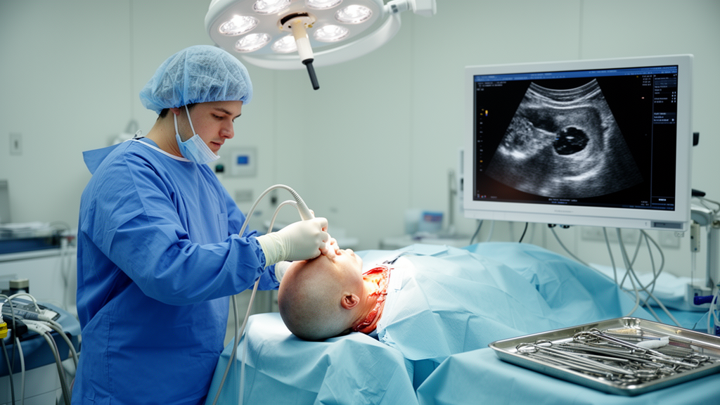
During the operation, the surgical team deployed intraoperative ultrasound using a high-frequency 7 to 12 MHz linear probe with sterile coupling gel, imaging in two orthogonal planes both before and after opening the dura. Baseline B-mode imaging — optimized by adjusting gain, depth, and focal zone to maximize lesion-to-parenchyma contrast — revealed a homogeneous, hyperechoic, poorly marginated area within the right frontal white matter, with preservation of the overlying cortical architecture. That pattern alone could easily be mistaken for a diffuse glioma, underscoring the diagnostic ambiguity the surgeons faced.
The real breakthrough came when the team administered sulfur hexafluoride microbubbles intravenously, followed by a saline flush, and recorded enhancement dynamics for 130 seconds using contrast-specific imaging software at a low mechanical index below 0.1. The contrast-enhanced ultrasound revealed a distinctive pattern: a delayed uptake phase beginning approximately 35 seconds after injection, peaking at around 55 to 60 seconds, with intense, vivid, and relatively long-lasting enhancement before washout occurred after two minutes. Notably, the deeper portions of the lesion took up contrast earlier and more pronouncedly than the superficial regions, suggesting a gradient in perfusion within the affected white matter.
Intraoperative flow cytometry of fresh tissue samples showed no abnormal immunophenotype consistent with lymphoma recurrence, while cerebrospinal fluid analysis revealed a non-specific increase in white blood cells and real-time polymerase chain reaction confirmed the presence of JCV DNA at 8,616.6 IU/mL. Histopathological examination of the biopsy tissue demonstrated extensive demyelination with loss of myelin but relative preservation of axons, numerous foamy macrophages containing myelin degradation products consistent with active demyelination, marked reactive astrocytic changes with enlarged atypical cells, and oligodendrocytes showing nuclear enlargement with a ground-glass appearance suggestive of viral cytopathic effect. Together with the JCV DNA detection, these findings confirmed a diagnosis of PML.
The patient was discharged on the fourth postoperative day in good neurological condition, walking independently, and subsequently began treatment with pembrolizumab — an immune checkpoint inhibitor that has shown promise in PML by restoring antiviral immune function. At the most recent follow-up, approximately eight months after diagnosis, he remains alive and in good condition with a Karnofsky performance status of approximately 90, a remarkable outcome given the typically dismal prognosis of the disease.
The ultrasound findings, while striking, must be interpreted with caution. The authors note that the delayed and relatively persistent enhancement pattern may reflect altered microvascular perfusion within demyelinated white matter rather than the neovascularization typical of high-grade tumors, which tend to show earlier and more heterogeneous contrast uptake. Radiation necrosis and other treatment-related changes may present with heterogeneous or peripheral enhancement patterns, adding further complexity. However, the authors emphasize that their interpretation remains speculative and that no definitive conclusions can be drawn from a single case regarding the underlying mechanisms or the reproducibility of these imaging characteristics.
From a practical standpoint, intraoperative ultrasound is widely used in brain tumor surgery but has seen limited application in multifocal or demyelinating disease, largely because such conditions are typically diagnosed through stereotactic needle biopsy rather than open surgery. In this case, the ultrasound and contrast-enhanced imaging contributed to targeting areas for biopsy, though it did not allow reliable differentiation between demyelinating and neoplastic pathology on its own. The authors position these techniques as supportive rather than diagnostic tools, potentially improving sampling accuracy when open biopsy is indicated. They describe the imaging features as hypothesis-generating and call for larger patient cohorts to determine whether specific ultrasound patterns can be consistently identified in PML and whether they may genuinely contribute to improved intraoperative decision-making. For now, the report stands as a rare and valuable first look at how a viral brain infection appears under the real-time eye of the operating room ultrasound.
Subject of Research: First intraoperative ultrasound characterization of progressive multifocal leukoencephalopathy
Article Title: Intraoperative contrast-enhanced ultrasound features of progressive multifocal leukoencephalopathy: a case report
Article References: Bonosi, L., & Tringali, G. (2026). Intraoperative contrast-enhanced ultrasound features of progressive multifocal leukoencephalopathy: a case report. Frontiers in Neuroimaging, 5, Article 1842218. https://doi.org/10.3389/fnimg.2026.1842218
Image Credits: AI Generated
DOI: 10.3389/fnimg.2026.1842218
Keywords: progressive multifocal leukoencephalopathy, intraoperative ultrasound, contrast-enhanced ultrasound, John Cunningham virus, neurosurgery, brain biopsy, demyelinating disease, mantle cell lymphoma, Intraoperative, contrast-enhanced, ultrasound, features
News Source: Kristina Jarvis. (October 17, 2026). Surgeons Capture First-Ever Ultrasound Images of a Rare Brain Infection That Mimics Cancer. Scienmag.