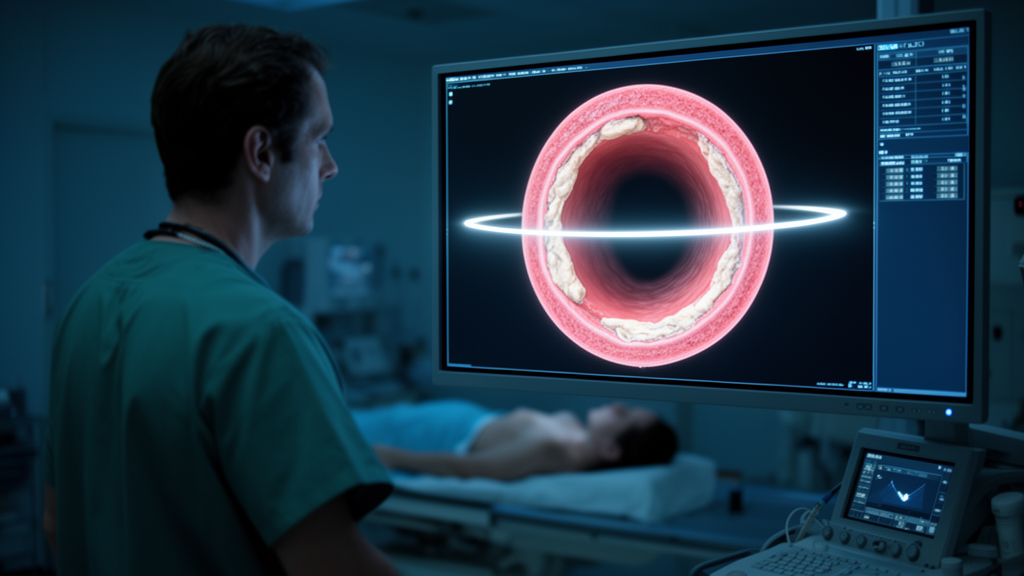
When a person survives an ischemic stroke, the danger is far from over. The brain has been injured, but the blood vessels feeding the heart may already be primed for the next catastrophe. A new study from Harbin Medical University in China, published in Frontiers in Cardiovascular Medicine, has now used one of the sharpest imaging tools in cardiology to show exactly what those vessels look like in patients arriving at the hospital with a heart attack after a previous stroke. The answer is unsettling: the coronary plaques in these patients are measurably more inflamed, more lipid-rich, and more structurally fragile than those in heart attack patients who never had a stroke.
The research team enrolled 1,050 patients with acute myocardial infarction, or AMI, who underwent optical coherence tomography, known as OCT, at the Second Affiliated Hospital of Harbin Medical University between September 2019 and December 2021. OCT is a near-infrared intravascular imaging technique that can resolve plaque structures with a precision of roughly 10 to 20 micrometers, allowing cardiologists to measure the thickness of the fibrous cap covering a plaque, map the extent of the lipid core, and identify inflammatory cells within the arterial wall. Of the enrolled patients, 193 had suffered an ischemic stroke before their heart attack, while 857 had no such history. To ensure that any differences in plaque characteristics were not simply the result of age, sex, or conventional risk factors, the investigators used 1:1 propensity score matching on variables including age, gender, hypertension, diabetes, dyslipidemia, smoking, and alcohol consumption, with a caliper width of 0.02. This produced a final cohort of 386 patients, 193 in each group, who were statistically comparable at baseline.
The technical definitions matter here. A lipid-rich plaque was defined as one with a lipid arc greater than 90 degrees on the OCT cross-section. The lipid core length was the longitudinal extent of that lipid-rich segment, and the lipid index was calculated as the lipid core length multiplied by the mean lipid arc, giving a composite measure of overall lipid burden. A thin-cap fibroatheroma, or TCFA, was defined as a plaque with a lipid pool arc of at least 180 degrees covered by a fibrous cap thinner than 65 micrometers at its thinnest point. TCFA is widely regarded as the OCT hallmark of the vulnerable plaque, the lesion most likely to rupture and trigger an acute coronary syndrome. Macrophages appeared on OCT as high-attenuation, signal-rich band-like or dot-like structures, typically accompanied by shadowing, marking active inflammation. Cholesterol crystals were identified as signal-rich linear structures within the necrotic core or adjacent to the fibrous cap.
The results for culprit lesions, the plaques that actually caused the heart attack, were striking. In the stroke group, 70.0 percent of culprit plaques were TCFAs, compared with 55.4 percent in the non-stroke group, a difference that was statistically significant at p = 0.003. Macrophage accumulation was present in 52.3 percent of culprit plaques in the stroke group versus 39.4 percent in the non-stroke group, p = 0.011. Cholesterol crystals were found in 35.2 percent versus 21.8 percent, p = 0.003. The lipid core was also longer in the stroke group, 15.3 plus or minus 7.4 millimeters versus 13.5 plus or minus 6.4 millimeters, p = 0.013, and the lipid index was higher, 3,465.0 plus or minus 2,135.8 versus 2,946.4 plus or minus 1,784.4, p = 0.011. Notably, there were no significant differences in minimal lumen area, percent area stenosis, fibrous cap thickness, or calcification, suggesting that the excess vulnerability was qualitative rather than purely a matter of vessel narrowing.
Perhaps even more important was what the investigators found in the non-culprit plaques, the lesions in other coronary vessels that had not yet caused an event but could do so in the future. The stroke group had a higher burden of multivessel disease, with 276 non-culprit plaques imaged across 367 coronary vessels, compared with 219 non-culprit plaques across 443 vessels in the non-stroke group. After adjusting for the clustering of multiple plaques within the same patient using generalized estimating equations, prior stroke remained independently associated with a higher prevalence of TCFA in non-culprit plaques, with an odds ratio of 2.08 and a 95 percent confidence interval of 1.15 to 3.74, p = 0.014. For culprit plaques, the corresponding odds ratio was 1.87, 95 percent confidence interval 1.23 to 2.84, p = 0.003. In other words, a history of ischemic stroke roughly doubled the odds that any given coronary plaque, whether it caused the current heart attack or not, would carry the OCT signature of vulnerability.
The clinical implications became clear during follow-up. Of the 386 matched patients, 382 completed 18 months of clinical follow-up, 191 in each group. The primary endpoint was major adverse cardiovascular events, or MACE, defined as cardiogenic death, non-fatal myocardial infarction, and ischemia-driven revascularization of target or non-target vessels. The stroke group experienced MACE at a rate of 8.9 percent versus 4.2 percent in the non-stroke group, p = 0.009, with a hazard ratio of 2.61, 95 percent confidence interval 1.28 to 5.34. Recurrent stroke occurred in 4.2 percent versus 1.6 percent, p = 0.019, hazard ratio 3.78, 95 percent confidence interval 1.23 to 9.30. Cardiogenic death was recorded in 5.8 percent versus 1.6 percent, p = 0.011, hazard ratio 3.55, 95 percent confidence interval 1.33 to 9.47. Kaplan-Meier analysis of the cumulative 18-month incidence confirmed significant differences between the groups for MACE, recurrent stroke, and cardiogenic death. There were no significant differences in recurrent myocardial infarction, readmission for angina or heart failure, revascularization, or bleeding events.
Why would a stroke leave such a signature in the coronary arteries? The authors point to the biology of systemic atherosclerosis. Ischemic stroke and AMI are both manifestations of the same underlying disease process, and prior studies have shown that 61.9 percent of patients who experienced a non-fatal ischemic stroke had evidence of coronary atherosclerotic plaque, while 25.7 percent exhibited coronary stenosis. The composite incidence of recurrent stroke, myocardial infarction, and vascular death within one year after a stroke has been documented at 24.7 percent. Even when patients are treated according to secondary prevention guidelines, roughly 25 percent still experience cardiovascular events. The new OCT findings suggest that prior stroke may act as a readily available clinical marker of a heightened systemic atherosclerotic burden and residual vascular risk, one that persists even after propensity matching on conventional risk factors.
The inflammatory dimension is particularly intriguing. The authors note that following an ischemic stroke, the body undergoes persistent inflammation, and recent work has shown that the cellular tissue of stroke patients contains mRNA profiles that can trigger inflammation and degrade the key structural components that prevent plaque rupture. Cholesterol crystallization, which was more prevalent in the culprit plaques of the stroke group, results from the accumulation of free cholesterol and the disruption of cholesterol homeostasis in macrophages. Excess esterified cholesterol and crystals released from dead foam cells into the extracellular space manifest as cholesterol crystals in coronary plaque, and their presence has been associated with coronary inflammation, plaque vulnerability, and poorer clinical outcomes within one year. The higher macrophage density observed in the stroke group is consistent with this pro-inflammatory milieu.
The study is not without limitations, and the authors are candid about them. The retrospective, single-centre design introduces selection bias, since only AMI patients with analyzable OCT images were included, which may limit generalizability. Despite propensity score matching, residual confounding from unmeasured factors such as inflammatory status and specific medication details cannot be entirely ruled out. Data regarding the subtypes, severity, and timing of prior stroke events were unavailable, precluding subgroup analysis. OCT evaluation was performed only at baseline, so the longitudinal evolution of plaque features could not be assessed. The authors also acknowledge that balance across all factors, including education level and inflammation, could not be achieved. These caveats mean the findings should be interpreted as an association rather than proof of causation.
Still, the message for clinical practice is hard to ignore. Large-scale imaging studies such as PROSPECT have shown that the positive predictive value of plaque morphological features like TCFA for future events is limited, roughly 15 to 25 percent, highlighting the necessity of integrating additional clinical characteristics to optimize risk stratification. A prior ischemic stroke may be exactly such a characteristic, a simple piece of clinical history that flags heart attack patients carrying a heavier load of OCT-defined high-risk plaques across multiple coronary vessels. The authors suggest that these patients may require more intensive secondary prevention strategies, although whether advanced imaging can improve risk stratification or clinical management requires further prospective investigation. For now, the study adds a granular, image-based dimension to a growing body of evidence that the heart and the brain are connected not just by the circulation, but by the fate of the plaques that line it.
Subject of Research: Cardiovascular Medicine
Article Title: Coronary plaque characteristics of acute myocardial infarction patients with and without previous ischemic stroke
Article References: Wang, Q., Dong, F., Fang, C., Hu, S., Jia, H., Pan, W., Li, L., Wu, J., He, L., Wang, Y., Chen, T., Yu, H., Dai, J., & Yu, B. (2026). Coronary plaque characteristics of acute myocardial infarction patients with and without previous ischemic stroke. Frontiers in Cardiovascular Medicine, 13, Article 1798730. https://doi.org/10.3389/fcvm.2026.1798730
Image Credits: AI Generated
DOI: 10.3389/fcvm.2026.1798730
Keywords: ischemic stroke, acute myocardial infarction, coronary plaque, optical coherence tomography, thin-cap fibroatheroma, plaque vulnerability, cardiovascular risk, atherosclerosis, Coronary, plaque, characteristics, acute
News Source: Cassandra Pierce. (October 17, 2026). Prior Stroke Leaves a Hidden Signature in the Coronary Arteries, OCT Study Reveals. Scienmag.