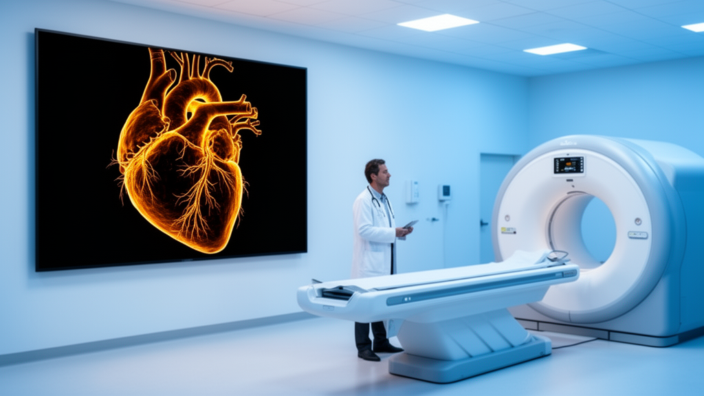
Every year, millions of patients undergo computed tomography angiography, one of the most powerful tools modern medicine has for peering inside the beating heart without opening the chest. The technique saves lives by revealing narrowed coronary arteries, dangerous plaques, and blood clots, but it carries a hidden cost: ionizing radiation. A new research project at The University of Texas at Arlington aims to change that equation, using artificial intelligence to dramatically cut the radiation dose patients receive during cardiac CT scans while preserving the diagnostic clarity physicians depend on.
The study, led by Mingwu Jin, professor of physics at UT Arlington, focuses on computed tomography angiography, or CTA, an advanced imaging test that noninvasively assesses coronary artery disease, the world’s leading cause of death. CTA combines a CT scan with iodine contrast dye injected into a patient’s vein to create detailed three-dimensional images of blood vessels and surrounding tissues throughout the body. Beyond the heart, doctors rely on the technique to detect aneurysms, narrowed or damaged vessels, blood clots, tumors, and a range of other conditions, which is precisely why reducing its radiation footprint could benefit so many people.
The project is funded by a three-year, $579,745 grant from the National Heart, Lung, and Blood Institute, part of the National Institutes of Health. It is titled Deep leaRning-bAsed reConstruction, or DRAC, for Multi-Phase Cardiac CT Angiography. The work targets a specific and increasingly important imaging protocol known as multi-phase coronary CTA, which captures images at multiple points during the heartbeat. By sampling the cardiac cycle repeatedly, the technique gives doctors far more information and helps reduce the blurring caused by the heart’s constant motion. The drawback is unavoidable physics: collecting images across multiple phases exposes patients to more radiation than a single-phase scan.
Lowering the radiation dose in medical imaging has been vigorously pursued due to imaging prevalence and the potential cancer risk of ionizing radiation, Jin explained. Although notable progress has been made by developing advanced reconstruction methods, the dose reduction potential is far from its limit. That statement captures the central tension of modern radiology: the same X-ray photons that produce exquisitely detailed images of the heart also carry a small but cumulative risk, and every reduction in dose must be earned without sacrificing the information clinicians need to make life-or-death decisions.
To understand how the UT Arlington team plans to earn that reduction, it helps to know what happens inside a CT scanner. The raw data collected during a scan are not images at all but measurements of X-ray attenuation from many angles. Reconstruction methods transform those measurements into the cross-sectional pictures doctors can interpret. One advanced approach is model-based iterative reconstruction, known as MBIR, an algorithm designed to generate high-quality cross-sectional images while potentially using less radiation. Rather than applying simple filtered back-projection, MBIR builds a mathematical model of the imaging process itself and iteratively refines the image until it best explains the measured data, suppressing noise in the process.
A second pillar of the project is 4D reconstruction, which adds the dimension of time to standard three-dimensional imaging. In cardiac imaging, this technique can capture clear views of fast-moving heart valves and coronary arteries while reducing motion artifacts, the ghostly streaks and blurs that arise when the heart moves between X-ray projections. By explicitly modeling motion over the cardiac cycle, 4D methods allow scanners to gather less data per phase while still producing images that look as though they were frozen at a single instant of the heartbeat.
Jin said that recent breakthroughs in deep learning provide an unprecedented opportunity to substantially lower radiation doses by combining MBIR with information learned from a large database of high-quality multi-phase coronary CTA images. The resulting 4D DRAC method would reconstruct a patient’s images using not only data from that individual scan, but also information learned from a much larger collection of images. In practical terms, the neural network acts as a kind of learned prior: it has seen what healthy and diseased coronary arteries look like in full-dose, high-quality scans, and it uses that statistical knowledge to fill in the details that a low-dose scan alone cannot resolve. This is the same broad principle that has revolutionized image denoising, super-resolution, and medical image synthesis across radiology, now aimed at one of the most technically demanding reconstruction problems in the field.
To test the hypothesis, Jin and his team will pursue three interlocking aims. First, they will build a synthetic patient library using computational human models based on retrospectively collected multi-phase coronary CTA data from real patients. Synthetic data allow the researchers to know the ground truth exactly, which is essential for training and validating deep-learning algorithms without exposing additional patients to scans. Second, they will develop three-dimensional and four-dimensional deep-learning methods that combine CT imaging physics with features learned from the high-quality patient data, ensuring the network respects the actual mathematics of X-ray attenuation rather than hallucinating plausible-looking anatomy. Third, they will determine the lowest radiation dose that can be used without compromising diagnostic accuracy, evaluating that accuracy with a computer model designed to mimic how a radiologist assesses an image, then validating the results through a small-scale study involving human observers.
The Mayo Clinic in Rochester, Minnesota, will provide multi-phase coronary CTA patient data for the study, giving the UT Arlington team access to clinically realistic images acquired on state-of-the-art scanners. That partnership matters because deep-learning reconstruction methods live or die by the diversity and quality of their training data; a network trained only on idealized images may fail when confronted with the variability of real patients, from differing heart rates and body habitus to calcium deposits and stents that scatter X-rays in complicated ways. By anchoring the synthetic library in retrospectively collected clinical data, the project aims to bridge the gap between algorithmic promise and bedside reality.
If the 4D DRAC methods succeed, the implications could extend well beyond the coronary arteries. Jin said the project could substantially lower the radiation dose needed for multi-phase coronary CTA, potentially improving its use in detecting the narrowing of coronary arteries and monitoring the progression of high-risk plaque, the unstable lesions most likely to trigger heart attacks. He noted that the methods could also be applied to other dynamic CT scans, which use a series of images to track changes over time, from perfusion studies of the brain to oncology follow-ups. And because the approach couples physics-based reconstruction with learned priors, it inherits the interpretability advantages of MBIR while gaining the pattern-recognition power of modern AI, a combination many researchers see as the most credible path to routine ultra-low-dose imaging.
The award also reflects the collaborative and educational dimension of the research. Graduate students Noor Rasel and Darnisha Langlais will work with Jin on the project, and the grant will allow him to hire undergraduate students in the summers, giving them hands-on opportunities to apply AI techniques to medical imaging. This award reflects the high quality of Dr. Jin’s research and the strength of his proposed ideas, said Alex Weiss, professor of physics and former department chair. I know a great deal of work goes into preparing a successful proposal of this kind. This is a significant achievement for Dr. Jin and a major benefit to our department. For the millions of patients who will undergo cardiac CT in the coming years, the hope is that the project’s most lasting contribution will be images just as sharp, diagnoses just as certain, and doses substantially smaller.
Subject of Research: AI-based low-dose reconstruction for multi-phase cardiac CT angiography
Article Title: UT Arlington study aims to cut radiation in heart scans
Article References: UT Arlington study aims to cut radiation in heart scans. (n.d.). Original publication
Image Credits: AI Generated
DOI: Not provided
Keywords: cardiac CT angiography, radiation dose reduction, deep learning, image reconstruction, coronary artery disease, University of Texas at Arlington, NIH funding, 4D reconstruction, model-based iterative reconstruction, medical imaging, machine learning, cardiovascular imaging
News Source: Ophelia Keating. (October 16, 2026). AI-Driven CT Reconstruction Aims to Slash Radiation in Heart Scans. Scienmag.