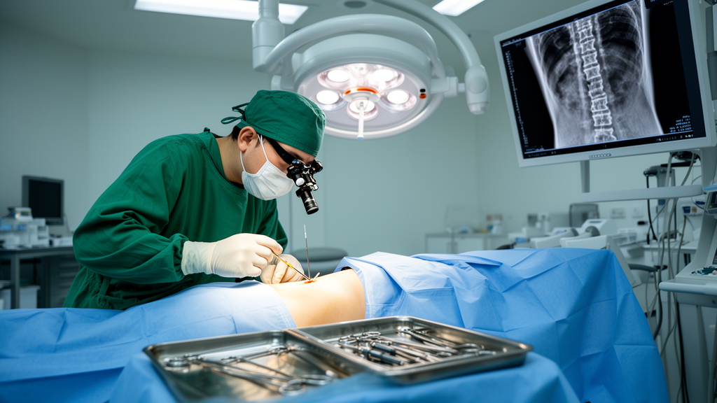
…
The dorsal root ganglion has long occupied a central position in pain neuroscience because it houses the cell bodies of primary afferent neurons that carry both somatic and visceral nociceptive information into the spinal cord. Genetic access to these neurons allows researchers to manipulate the molecular machinery of pain signaling with a precision that systemic pharmacology cannot match. Yet the technical difficulty of reaching the ganglion surgically has historically constrained how ambitious such experiments can be. The new protocol addresses this bottleneck by rethinking the anatomy of the surgical approach itself, and the details of that rethinking deserve closer examination because they illuminate why conventional methods have been so problematic.
Traditional intraganglionic injection techniques typically demand a wide laminectomy, in which substantial portions of the vertebral arch are removed to expose the underlying ganglion. In some protocols, the facet joints themselves are excised to widen the surgical corridor. While this generous exposure certainly makes the ganglion easier to see and puncture, it comes at a biomechanical cost. The facet joints, together with the intervertebral discs and ligaments, form the functional spinal unit that stabilizes the column while preserving flexibility. Removing even one facet complex can alter load distribution across adjacent levels, and multi-level resection compounds this effect. In small animals such as mice, whose vertebrae are only a few millimeters across, the margin between adequate exposure and destabilizing destruction is narrow. Instability in turn promotes compensatory postural changes, altered gait, and mechanical allodynia that can confound the very pain phenotypes an experiment is designed to measure.
Tissue adhesion presents a second, less obvious obstacle. Following extensive bony and muscular dissection, fibrotic scar tissue forms between the overlying paraspinal muscles and the denuded bone. If a second surgery is required at the same segment, whether to inject a different viral construct, to deliver a second vector weeks after the first, or to implant an imaging window, the surgeon must dissect through this scarred plane. Each round of re-dissection increases bleeding, obscures landmarks, and raises the risk of injuring the nerve roots and ganglia that the experiment intends to study. The adhesion problem effectively caps the number of sequential interventions an animal can tolerate, which limits longitudinal experimental designs that are increasingly valued in chronic pain research, where the same animal must be followed across the weeks needed for disease progression or gene expression to evolve.
The multi-segment problem is a third constraint. Many questions in pain neuroscience cannot be answered by manipulating a single ganglion. Convergence of somatic and visceral afferent inputs onto shared spinal projection neurons, the segmental organization of referred pain, and the coordinated sensitization of neighboring dermatomes all require simultaneous or sequential access to several contiguous ganglia. The lumbar levels L3 through L6 in the mouse are particularly relevant because they innervate the hindlimb and pelvic viscera, making them the natural target set for studies of somatic-visceral crosstalk. Conventional single-ganglion protocols, scaled up by repeating the exposure at each level, multiply the surgical burden and the associated instability and adhesion problems. A method that permits several ganglia to be reached through one limited exposure therefore changes the practical calculus of experimental design.
The anatomical foundation of the new approach rests on identifying reliable landmarks in a region where they are scarce. Mouse lumbar vertebrae are small, morphologically similar to one another, and variable in number because of lumbosacral transitional vertebrae, a well-documented skeletal variant in laboratory strains. Miscounting segments is consequently one of the most common silent errors in spinal surgery in mice, and an injection intended for L4 may instead land in L3 or L5, invalidating the interpretation of downstream anatomical and functional data. The counting scheme described here exploits the V-shaped white lines visible on the spinous processes, bony features that can be identified through the surgical field without additional imaging. Counting caudal to rostral, from a confidently identified caudal anchor toward the target levels, reduces cumulative error compared with counting in the opposite direction, because the caudal landmarks are reportedly more distinctive.
Before committing to a surgical target, the investigators mapped which ganglia actually innervate which peripheral territories, using transgenic mice that express yellow fluorescent protein in sensory neurons. The resulting segmental map carries practical consequences for experimental design. Fibers from L1 and L2 do not contribute to the sciatic nerve, so experiments on hindlimb somatosensation that rely on sciatic-dependent behaviors should target L3 through L5, which collectively give rise to that nerve. L6 contributes predominantly to the pudendal nerve, with minor contributions from L5, which matters for studies of pelvic visceral nociception. These findings also highlight a known pitfall in cross-species extrapolation: segmental contributions differ between mouse and rat, and published mouse maps themselves show discrepancies that are complicated by lumbosacral skeletal variants. An in-house validation of the map in the specific strain being used is therefore not pedantry but a safeguard against misattributing behavioral outcomes to the wrong segments.
The key surgical insight is that the spatial relationship between each lumbar DRG and the facet joints of the neighboring vertebrae is stereotyped enough to serve as a navigation aid. Anatomical dissection demonstrated that drilling at the base of the inferior articular process of the vertebra cranial to the target ganglion provides a direct access route to the L3 through L6 DRGs. The inferior articular process is a small bony projection that forms the lower half of the facet joint, and its base sits in a predictable relationship to the ganglion below. By drilling a small window at this location rather than removing the entire lamina or joint, the surgeon preserves the majority of the posterior column while still reaching the ganglion with a fine injection needle. The approach is conceptually similar to minimally invasive foraminotomy techniques in human spine surgery, where limited bony windows replace wide exposures, adapted here to the millimeter scale of the mouse.
Preserving the facet joints addresses the stability problem, but a remaining challenge is the movement of the ganglion itself during injection. The DRG is not fixed in place; it moves with each respiratory cycle and heartbeat as the surrounding soft tissues shift. In a needle as fine as those used for intraganglionic microinjection, even sub-millimeter relative motion can lacerate neuronal cell bodies, turning a delivery procedure into an injury model. Conventional spinal suspension holders that lift the animal by the spinous processes reduce but do not eliminate this motion, particularly across the extended operative field needed for multi-segment work, where residual segment-level soft-tissue shift persists. The custom temporary spinal stabilizer described in the protocol is designed to immobilize the relevant segment locally during the injection moment, then be removed, avoiding the chronic alterations associated with permanent hardware.
Validation proceeded along two complementary axes. The first was behavioral: mice receiving injections at one to as many as eight ganglia were followed serially after surgery. Functional disturbances appeared transiently, consistent with the expected sequelae of any spinal surgery, but most resolved by postoperative day 21. This timeline matters for experimental planning, because it defines a window after which baseline behavior can be assumed to have returned before experimental manipulations or pain models are initiated. It also provides a benchmark against which future users of the protocol can judge whether their surgical technique is producing acceptable levels of transient deficit or an abnormal rate of lasting impairment.
The second axis was functional confirmation of transduction. Viral expression alone does not guarantee that transduced neurons remain physiologically competent, so the investigators turned to in vivo calcium imaging using the genetically encoded indicator jGCaMP8s. In transduced animals, air-puff stimulation of the periphery evoked robust calcium responses in the DRG, demonstrating that the transduced sensory neurons could still detect peripheral stimuli and transduce them into neural activity. Wild-type mice, which lack the indicator, showed no such signals, serving as negative controls and confirming that the measured fluorescence reflected genuine neural activity rather than imaging artifact. This combination of anatomical targeting, behavioral recovery, and functional readout forms a closed loop of validation that individual pieces of evidence alone cannot provide.
The broader significance of the method lies in what it enables rather than in any single result. Multi-segmental access to L3 through L6 ganglia in the same animal opens the door to experiments in which different segments receive different constructs, allowing within-animal comparisons that reduce the confounding of inter-individual variability. Repeated access without extensive adhesion supports longitudinal gene manipulation schedules matched to the slow time course of chronic pain models. The compatibility with in vivo calcium imaging means that the same animals can be followed functionally over time, connecting molecular manipulation to circuit-level activity and behavioral output within a single cohort. For the study of somatic-visceral pain crosstalk, where convergent afferent signaling across multiple segments is thought to be central, these capabilities address precisely the experimental constraints that have made such studies difficult to execute cleanly.
As with any surgical protocol, generalization will require independent replication across laboratories, and the technique demands the microsurgical skill and anatomical familiarity that spinal injection work in mice has always required. The transient behavioral disturbances, while resolving in most animals, underscore that the procedure is not benign, and appropriate controls and monitoring remain essential. Nevertheless, by anchoring the approach in validated anatomical landmarks, minimizing the destruction of load-bearing structures, and pairing delivery with functional verification, the workflow offers a coherent template for DRG-targeted experiments that is more scalable and more repeatable than the conventional alternatives it seeks to replace.
Subject of Research: …
Article Title: A facet joint landmark-guided approach for multi-segmental DRG injection with limited bone removal in mice
Article References: A facet joint landmark-guided approach for multi-segmental DRG injection with limited bone removal in mice. (n.d.). https://doi.org/10.3389/fneur.2026.1956927
Image Credits: AI Generated
DOI: 10.3389/fneur.2026.1956927
Keywords: facet, joint, landmark-guided, approach, multi-segmental, injection, limited, bone, removal, mice, scientific research
News Source: Ophelia Keating. (October 16, 2026). New Landmark-Guided Surgery Lets Scientists Inject Genes Into Multiple Spinal Nerve Hubs at Once. Scienmag.