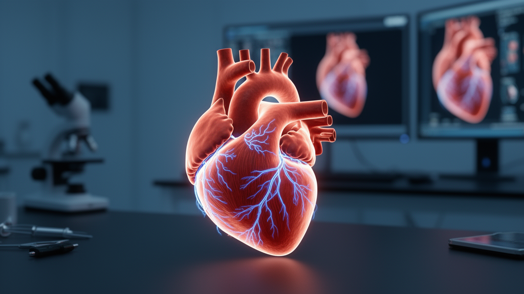
Sudden cardiac death remains one of medicine’s most devastating and unpredictable outcomes, claiming millions of lives each year. A new review published in Frontiers in Cardiovascular Medicine argues that the field’s reliance on a single, decades-old metric—left ventricular ejection fraction—is holding back accurate risk prediction, and that personalized computational heart models, a medical embodiment of the digital twin concept, offer a way forward. The authors, cardiologists from Capital Medical University Affiliated Anzhen Hospital in Beijing, systematically lay out how a patient’s own cardiac magnetic resonance imaging can be transformed into a beating virtual heart capable of revealing hidden electrical instability before it strikes.
The scale of the problem is staggering. Ventricular fibrillation and ventricular tachycardia account for the vast majority of sudden cardiac deaths, which occur roughly 350,000 times annually in Europe and North America and about 540,000 times each year in China. In adults over 35, coronary artery disease is responsible for more than 80 percent of these events. Most survivors of a myocardial infarction develop scar tissue that replaces dead heart muscle, and it is this scar—particularly its heterogeneous border zone, where surviving myocytes intermingle with collagen deposits—that creates the electrical Short-circuit pathways for re-entrant ventricular arrhythmias.
Yet the clinical tools currently used to identify who will succumb to these arrhythmias are strikingly limited. Risk stratification in routine practice hinges on ejection fraction, a measure of pumping efficiency that captures nothing about the underlying electrical substrate. The consequences are visible in the prophylactic use of implantable cardioverter-defibrillators, which are both under-implanted in patients with preserved function who nonetheless die suddenly, and over-implanted in patients whose devices never fire. Invasive electrophysiological studies, the traditional gold standard for inducibility testing, carry procedural risks and cannot sample the whole heart; the landmark MADIT II analysis showed their predictive value for future events is limited. Even late gadolinium enhancement cardiac magnetic resonance, which beautifully maps scar architecture, cannot reveal conduction velocity, refractory behavior, or decremental conduction within scar channels—the very properties that determine whether a re-entrant circuit can form and sustain itself.
Personalized cardiac modeling closes this gap by integrating multi-modal data into a physics-based computational replica of the patient’s ventricles. The pipeline begins with late gadolinium enhancement MRI, where pixels exceeding signal intensity thresholds—typically five standard deviations above normal myocardium—are classified as scar. Segmentation techniques, whether semi-automatic workflows with manually placed feature points or emerging fully automatic deep-learning tools, convert the images into three-dimensional geometric models and finite element meshes. Infarct regions are classified into a dense scar core and a partially infarcted border zone, and rule-based algorithms assign the helical orientation of myocardial fibers that governs anisotropic electrical conduction through the ventricular wall.
Onto this anatomical scaffold, the modelers graft cellular electrophysiology. Established human ventricular cell models—including the ten Tusscher–Noble–Panfilov model, the O’Hara–Rudy model, the Grandi–Bers model, and the more recent ToR-ORd refinement—describe the choreography of ion currents that shapes each action potential, from the fast sodium current driving depolarization to the L-type calcium current and delayed rectifier potassium currents sustaining the plateau. In the infarct border zone, these conductances are downregulated to reflect experimentally documented remodeling: a widely cited parameterization applies reductions of 62 percent in sodium current, 69 percent in L-type calcium current, and 70 to 80 percent in delayed rectifier potassium currents, producing prolonged action potentials and reduced depolarization velocity. The review is candid about the limitations of this approach—the scaling factors derive largely from canine patch-clamp data extrapolated to human models, applied uniformly rather than across the true spatial gradients of the border zone, and omit features such as late sodium current upregulation and fibroblast coupling.
At the tissue level, electrical propagation is typically governed by the monodomain reaction–diffusion equation with anisotropic conductivity tensors. Dense scar is treated as electrically non-conductive, while border-zone transverse conductivity is reduced—by 90 percent in the canonical calibration—to mimic connexin-43 remodeling, yielding a normal longitudinal conduction velocity of about 0.6 meters per second. Alternative strategies represent diffuse microfibrosis as stochastic removal of intercellular links near a percolation threshold, or use Perlin-noise texture generation and homogenization techniques to preserve the fine-scale architecture of collagen strands that influences conduction slowing and spiral wave anchoring. The review stresses a critical caveat: different static fibrosis representations applied to the same imaging data can yield markedly different simulated rotor dynamics, and static conductivity maps are fundamentally incapable of reproducing the rate-dependent conduction block that characterizes diseased tissue.
The predictive climax of the workflow is the virtual electrophysiological study. Programmed electrical stimulation is applied at 19 standardized sites across the computational heart—a baseline pacing train followed by extrastimuli of progressively shorter coupling intervals—mimicking the catheter laboratory protocol. If the simulation induces ventricular tachycardia or fibrillation, the patient’s digital twin is flagged as susceptible to sudden death. In a pivotal validation study, this approach correctly predicted arrhythmic outcomes in post-infarction patients and outperformed ejection fraction, scar volume, gray zone volume, and left ventricular mass. Complementary metrics such as the Global Heterogeneity Score, computed from pixel-level signal intensity variation within scar, and the VITA framework, which automatically detects re-entrant pathways and computes their round-trip times, have shown independent associations with appropriate ICD therapy and with recurrence risk after catheter ablation.
Clinical validation is accumulating but incomplete. In hypertrophic cardiomyopathy, personalized models integrating late gadolinium enhancement and T1 mapping have predicted ventricular arrhythmia risk significantly better than guideline-based approaches. Meanwhile, the recent CMR GUIDE randomized trial of 353 patients with mildly to moderately reduced ejection fraction and CMR-detected scar found that ICD implantation did not significantly reduce the composite primary endpoint, though sudden cardiac death itself occurred less frequently in the ICD group. The authors interpret this as evidence that scar carries clinically relevant arrhythmic information that ejection fraction misses, but that scar-based selection alone still cannot capture the full electrophysiological heterogeneity—precisely the domain where dynamic, mechanism-based simulation may excel.
Significant hurdles stand between these models and routine practice. Whole-heart simulations involve millions of grid nodes and dozens of coupled differential equations spanning time scales from milliseconds to months, demanding high-performance computing. Segmentation remains partially manual and operator-dependent; ionic parameters rely on population averages rather than individual measurements; and no universally accepted gold-standard biomarker exists for validating sudden death predictions. The review points toward a future of multi-scale electromechanical coupling, deep-learning surrogate models that infer infarct anatomy directly from 12-lead electrocardiograms, calibration against electroanatomical mapping data, and treatment decision platforms that simulate ablation lines or drug effects in silico before the patient is touched. Interdisciplinary advances—from GPU acceleration to high-density microelectrode arrays—will be needed to complete the paradigm shift the authors envision: from passive treatment of arrhythmias after they strike, to active, personalized prevention grounded in a digital replica of each patient’s own heart.
Subject of Research: Personalized computational cardiac modeling for predicting sudden cardiac death risk after myocardial infarction
Article Title: Advances and challenges in personalized cardiac modeling and simulation for predicting the risk of sudden death from malignant arrhythmias in post-myocardial infarction
Article References: Zhao, E., Cheng, L., Liu, H., Wang, Z., & Wu, Y. (2026). Advances and challenges in personalized cardiac modeling and simulation for predicting the risk of sudden death from malignant arrhythmias in post-myocardial infarction. Frontiers in Cardiovascular Medicine, 13, Article 1879657. https://doi.org/10.3389/fcvm.2026.1879657
Image Credits: AI Generated
DOI: 10.3389/fcvm.2026.1879657
Keywords: sudden cardiac death, cardiac digital twin, myocardial infarction, ventricular arrhythmia, late gadolinium enhancement MRI, ventricular tachycardia, computational modeling, cardiac electrophysiology, implantable cardioverter-defibrillator, risk stratification, myocardial fibrosis, precision medicine
News Source: Ophelia Keating. (October 15, 2026). Digital Twin Hearts: How Personalized Cardiac Simulations Could Predict Sudden Death After a Heart Attack. Scienmag.