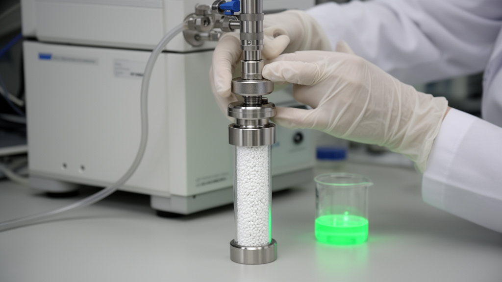
Fluorescently labeled proteins sit at the heart of modern biochemistry, powering everything from cellular imaging assays to the development of antibody-drug conjugates. Yet behind every glowing image lies a stubborn analytical problem: once a protein has been chemically decorated with a dye, researchers need a fast and reliable way to confirm what is actually in the reaction tube. A reaction mixture typically contains three species at once — the unmodified protein, the dye-conjugated protein, and leftover free dye — and teasing them apart has traditionally required either denaturing gel electrophoresis, which destroys the intact protein, or laborious peptide mapping, which digests the protein into fragments and sacrifices the big picture. Now, a team of Japanese researchers led by Shunsuke Yamazaki, Kazutoshi Takahashi, and Yutaka Matsuda of Ajinomoto Co., Inc. has reported a practical solution built on an unexpected chromatographic workhorse: the polyphenyl reversed-phase column.
The study, published in the open-access journal Discover Chemistry, describes a reversed-phase high-performance liquid chromatography method with photodiode-array detection, abbreviated RP-HPLC-PDA, that can monitor all three components of a protein-dye labeling reaction in a single intact-protein run. The test subjects were human serum albumin (HSA) and bovine serum albumin (BSA), two closely related blood proteins that the team had previously modified using an elegant enzymatic strategy. The work addresses a genuine gap in the bioconjugation toolkit, because intact-level chromatographic separation of native protein conjugates has long been more art than science, with analysts forced to guess which column chemistry might tame a given protein-dye combination.
The labeling workflow at the center of the study relies on Escherichia coli lipoate ligase A, or LplA, an enzyme that naturally attaches lipoic acid to specific lysine residues within a recognition tag. What makes LplA especially valuable for bioconjugation is that it can be coaxed into installing synthetic analogs, such as 8-azidooctanoic acid, onto native proteins without any genetic engineering. The azide group then serves as a chemical handle for strain-promoted azide-alkyne cycloaddition, a copper-free click reaction, with a dibenzocyclooctyne-bearing fluorescent probe such as DBCO-Cy3. The result is a tag-free modification strategy that operates directly on proteins in their native form — a significant advantage when the target protein cannot easily be redesigned or recombinantly expressed with an added tag.
With the chemistry established in their earlier work, the researchers turned to the analytical bottleneck. After LplA-mediated azide installation and DBCO-Cy3 click labeling, the sample contains unmodified protein, Cy3-labeled protein, and residual free dye. Sodium dodecyl sulfate-polyacrylamide gel electrophoresis with fluorescence imaging can confirm that labeling occurred, but it cannot deliver an intact chromatographic separation of the conjugated and unconjugated species. Peptide mapping offers residue-level detail but demands enzymatic digestion and is poorly suited to rapid screening. The team therefore sought a single, simple, intact-level method capable of tracking protein, protein-dye conjugate, and free dye simultaneously — a capability that would dramatically accelerate reaction optimization and sample selection.
The heart of the new paper is a systematic column screen. Using an Agilent 1260 HPLC system with a binary gradient pump, temperature-controlled column compartment, and photodiode-array detector, the researchers compared four reversed-phase column formats under identical conditions: a C18 column (XBridge BEH Shield RP18), a C8 column (YMC-Triart C8), a polystyrene/divinylbenzene column (Sepax Proteomix RP-1000), and a polyphenyl column (BioResolve RP mAb polyphenyl). The mobile phases were 0.1% trifluoroacetic acid in water and in acetonitrile, a classic ion-pairing system for protein chromatography, and the column temperature was held at 70 degrees Celsius to promote elution and sharpen peaks for intact proteins. Detection was performed at 280 nanometers, where proteins absorb, and at 570 nanometers, where Cy3 absorbs — a dual-wavelength arrangement that lets the analyst read protein and dye signals independently in the same run.
The results of the screen were strikingly divergent. The C8 column failed to retain intact HSA at all, with the protein signal eluting near the void volume and merging with the free dye — a category the authors labeled “not captured.” The C18 column showed the opposite pathology: severe late or incomplete elution, consistent with excessive hydrophobic retention under the screening gradient. The polystyrene/divinylbenzene column offered only partial retention, producing a large void-volume component and a broad late-eluting signal that was not useful for intact monitoring. Only the BioResolve RP mAb polyphenyl column delivered what the authors call “effective elution”: intact protein species eluted cleanly, free DBCO-Cy3 appeared as a separate peak, and the unmodified and Cy3-labeled protein forms could be distinguished by combining the 280 and 570 nanometer signals.
Importantly, the authors are careful about what this comparison does and does not prove. The four columns differed not only in bonded-phase chemistry but also in pore size, particle size, support material, and particle morphology — the polyphenyl column, for instance, features wide 450-angstrom pores and superficially porous 2.7-micrometer particles, while the PS/DVB column used 1000-angstrom pores with 5-micrometer fully porous particles. The improved performance of the polyphenyl format therefore reflects the combined effect of its wide-pore architecture, superficially porous design, and aromatic surface chemistry, and cannot be attributed to polyphenyl bonding alone. The screening conditions were deliberately common and non-optimized, making the exercise a practical starting-point exercise rather than a controlled study of stationary-phase selectivity. This kind of methodological honesty is refreshing in a field where column vendors often market surface chemistry as a magic bullet.
As a demonstration application, the team applied the method to compare Cy3-labeled HSA and BSA, two serum albumin homologs with closely related three-dimensional folds. Despite their structural kinship, the two proteins had shown markedly different apparent labeling efficiencies in the earlier substrate-panel study: HSA yielded an apparent Cy3-to-protein signal ratio of roughly 3, while BSA yielded roughly 1 under identical conditions. The intact RP-HPLC-PDA data confirmed this protein-level difference, but the authors emphasize that the method cannot identify which lysine residue carries the dye. Peak areas at 570 nanometers represent an apparent labeling readout rather than an absolute site-occupancy measurement, and residue-level assignment still requires peptide mapping or other site-resolved mass spectrometric approaches.
To explore why HSA outperformed BSA, the researchers conducted an exploratory lysine microenvironment analysis using solvent-accessible surface area values extracted from crystal structures of the two proteins. A simple exposure filter proved insufficient: BSA actually contains more lysines above the 160-square-angstrom exposure threshold than HSA, yet labeled less efficiently. Digging deeper, the team examined the immediate sequence neighbors of high-exposure lysines, reasoning that LplA-mediated labeling proceeds through an adenylated 8-azidooctanoate intermediate and that nearby basic residues might influence productive labeling. In HSA, high lysine exposure and a neighboring basic residue frequently coincided — for example, in His-Lys-Thr at positions 465 to 467 and Gly-Lys-Lys at positions 572 to 574 — whereas in BSA these features were more often distributed across separate regions. This pattern offers a plausible, hypothesis-generating explanation for the labeling difference, though the authors stress that it is a prioritization framework for future peptide mapping, not a mechanistic proof or a site-assignment rule.
The study comes with clearly stated limitations that researchers should heed before adopting the method. It was not validated as a quantitative assay — detection limits, quantification limits, and precision metrics were not systematically determined — so it is best used for qualitative or semi-quantitative monitoring. The lysine analysis rests on static structural models and ignores electrostatic potential, side-chain dynamics, steric accessibility to the enzyme, and conformational flexibility, any of which could shape real labeling behavior. Within those boundaries, however, the contribution is genuinely useful: the polyphenyl RP-HPLC-PDA platform gives bioconjugation labs a fast front-end screening tool for verifying that a protein is labeled, confirming that free dye has been removed, and comparing apparent labeling across substrates — all without digesting or denaturing the sample. Samples showing promising intact-level profiles can then be routed to peptide mapping or intact mass spectrometry for definitive characterization. For a field racing to build cleaner antibody-drug conjugates, imaging probes, and functionalized biologics, a reliable first look at the intact conjugate may prove to be exactly the accelerant the workflow needed.
Subject of Research: Intact reversed-phase HPLC analysis of Cy3-labeled serum albumin conjugates prepared by LplA-mediated tag-free protein modification
Article Title: Polyphenyl reversed phase chromatography separates Cy3 labeled serum albumins from unmodified proteins and free dye
Article References: Yamazaki, S., Takahashi, K., & Matsuda, Y. (2026). Polyphenyl reversed phase chromatography separates Cy3 labeled serum albumins from unmodified proteins and free dye. Discover Chemistry, 3(1), Article 438. https://doi.org/10.1007/s44371-026-00902-3
Image Credits: AI Generated
DOI: 10.1007/s44371-026-00902-3
Keywords: RP-HPLC, polyphenyl column, LplA, protein labeling, serum albumin, Cy3, click chemistry, bioconjugation, photodiode-array detection, intact protein analysis, HSA, BSA
News Source: Bethany Barker. (October 15, 2026). Polyphenyl Columns Emerge as a Practical Route to Intact Analysis of Fluorescent Protein Conjugates. Scienmag.