A drug designed to put patients safely to sleep on the operating table has become the focus of an urgent addiction investigation. Etomidate, a short-acting anesthetic that acts selectively on the brain’s main inhibitory receptor, the GABA type A receptor, has in recent years emerged as a substance of misuse in parts of Asia, and health authorities have struggled to understand why. Now a team of Chinese neuroscientists has produced one of the first detailed maps of what chronic etomidate misuse does to the human brain, combining electrical recordings, functional brain scans, and structural imaging in a single case-control study. The results, published in BMC Medicine, point to a specific prefrontal-to-parietal circuit whose breakdown appears to drive the intense craving that keeps users locked in the cycle of dependence.
The research was led by Junwei Zheng, Taoyu Wu, and colleagues working with Jie Shi and Yan Sun at Peking University’s National Institute on Drug Dependence, together with Sizhi Ai at Guangzhou Medical University. Between June 2023 and October 2024, the team recruited right-handed men aged 18 to 45 who met the diagnostic criteria for etomidate use disorder under the fifth edition of the Diagnostic and Statistical Manual of Mental Disorders, alongside age-matched healthy male controls. All participants were assessed in Guangzhou, and the study received ethics approval from Peking University Health Science Center with written informed consent from every participant. By restricting the sample to men within a defined age band, the investigators controlled for the hormonal and anatomical variability that often complicates addiction neuroimaging studies.
The experiment centered on a cue-reactivity paradigm, a standard but powerful technique in addiction science. Participants viewed images associated with the drug alongside neutral pictures, while researchers measured how the brain responded to each category. Because craving unfolds on multiple timescales, from milliseconds to seconds, the team deliberately paired two complementary imaging technologies. Electroencephalography captures the brain’s electrical activity with millisecond precision through electrodes on the scalp, while functional magnetic resonance imaging tracks changes in blood oxygenation that mark where neural activity is concentrated over seconds. Independent cohorts completed each modality, allowing the researchers to test whether the electrical and hemodynamic signatures of craving converged on the same circuitry.
The EEG findings were striking. When people with etomidate use disorder viewed drug-related images, their brains produced markedly amplified late positive potentials, a slow electrical wave that emerges roughly 300 milliseconds after a stimulus and peaks over frontocentral electrode sites. The late positive potential has long been treated as an index of motivated attention: the brain flagging something as salient and worth allocating resources to. In this study, the size of the etomidate-related signal correlated directly with participants’ subjective ratings of craving, measured with a desire-for-drug questionnaire. In other words, the brains of dependent users were not passively registering the pictures; they were actively and involuntarily magnifying their importance, and the degree of that amplification tracked how strongly each individual wanted the drug at that moment.
The fMRI data told a spatially precise story that converged with the electrical one. Drug cues triggered hyperactivation in two regions: the temporoparietal junction, an area at the intersection of temporal and parietal cortex involved in attentional orienting and salience detection, and the ventrolateral prefrontal cortex, a region classically associated with cognitive control and the regulation of impulses. Hyperactivation of the ventrolateral prefrontal cortex might sound like a signature of stronger control, but the connectivity analysis revealed something more troubling about what that activation was actually doing.
To move beyond simple co-activation maps, the researchers applied dynamic causal modeling, a computational framework that estimates the direction and sign of influences between brain regions rather than merely noting which areas light up together. The signed connectivity parameters exposed a critical abnormality: in the etomidate group, the ventrolateral prefrontal cortex exerted abnormal inhibitory modulation over the temporoparietal junction, and crucially, the strength of that inhibition was linked to craving severity. Participants with weaker inhibitory modulation reported higher subjective craving. This finding reframes the role of the prefrontal cortex in the disorder. Rather than simply failing to engage, the control system appears to be miswired, its top-down signal to the attention-grabbing temporoparietal junction weakened precisely when it would be needed most to disengage from drug-related cues.
Structural MRI added a third and sobering layer of evidence. Using surface-based morphometry to measure cortical thickness across the entire brain, the team identified widespread thinning among individuals with etomidate use disorder, spanning prefrontal, parietal, occipitotemporal, and limbic regions. Cortical thinning in these territories has been reported in other substance use disorders and is generally interpreted as reflecting chronic neuroadaptation, whether through synaptic pruning, glial changes, or loss of dendritic complexity. That etomidate, a drug once considered relatively benign because of its rapid clearance and organ-sparing profile, is associated with structural changes across such a broad cortical expanse underscores that apparent pharmacological safety in the anesthetic context says little about safety under the repeated, escalating exposure patterns of misuse.
Etomidate’s pharmacology makes the findings biologically coherent. The drug is a GABA-A receptor positive allosteric modulator, and the GABAergic system is deeply intertwined with the reward and executive circuits implicated in every major addiction. Chronic exposure to substances that enhance inhibitory signaling forces the brain to recalibrate, and those recalibrations plausibly extend to the prefrontal networks that govern attention and impulse regulation. The study’s convergent design strengthens this interpretation: an electrical marker of exaggerated salience attribution, a hemodynamic marker of cue-driven hyperactivation, a connectivity marker of impaired top-down inhibition, and a structural marker of cortical compromise all point toward the same VLPFC-TPJ circuit as the neural seat of etomidate craving.
What elevates the study from descriptive to clinically consequential is the target it nominates. Non-invasive brain stimulation techniques, including transcranial magnetic stimulation and transcranial direct current stimulation, can modulate prefrontal activity with growing precision, and randomized trials in other addictions have already explored the ventrolateral prefrontal cortex as a stimulation site. If weakened VLPFC-to-TPJ inhibition is genuinely causal in maintaining craving, then restoring that inhibitory modulation becomes a testable therapeutic hypothesis, one that could be evaluated with stimulation protocols, neurofeedback, or even pharmacological agents that strengthen prefrontal function. The authors position the VLPFC-TPJ circuit explicitly as a promising neuromodulation target for treating etomidate use disorder, a framing that could accelerate translational work in a field that currently has no approved pharmacological treatment.
The study’s limitations invite caution and future work. The cohort was restricted to right-handed men, so the findings cannot yet be generalized to women or to patterns of use in other populations, and the case-control design cannot establish whether the cortical thinning preceded drug use or resulted from it. Longitudinal follow-up, ideally beginning before dependence develops, will be needed to disentangle cause from consequence. Still, as a first multimodal portrait of an emerging and poorly understood form of addiction, the work delivers something rare: a mechanistically specific, biologically grounded, and clinically actionable account of craving. At a moment when etomidate misuse is escalating globally, that combination may prove exactly what public health responses have been missing.
Subject of Research: Neural mechanisms of cue-induced craving and cortical structural changes in etomidate use disorder
Article Title: Multimodal neuroimaging of cue-induced craving and cortical alterations in etomidate use disorder
Article References: Multimodal neuroimaging of cue-induced craving and cortical alterations in etomidate use disorder. (n.d.). https://doi.org/10.1186/s12916-026-05253-5
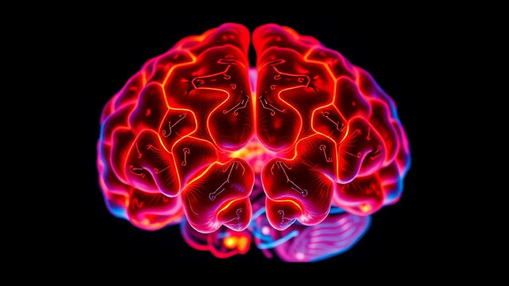
Image Credits: AI Generated
DOI: 10.1186/s12916-026-05253-5
Keywords: etomidate use disorder, drug craving, cue reactivity, EEG, fMRI, late positive potential, ventrolateral prefrontal cortex, temporoparietal junction, dynamic causal modeling, cortical thickness, GABA-A receptor, addiction neuroscience
News Source: Colin Clarke. (October 14, 2026). Brain Imaging Reveals How an Anesthetic Drug Hijacks Craving Circuits. Scienmag.