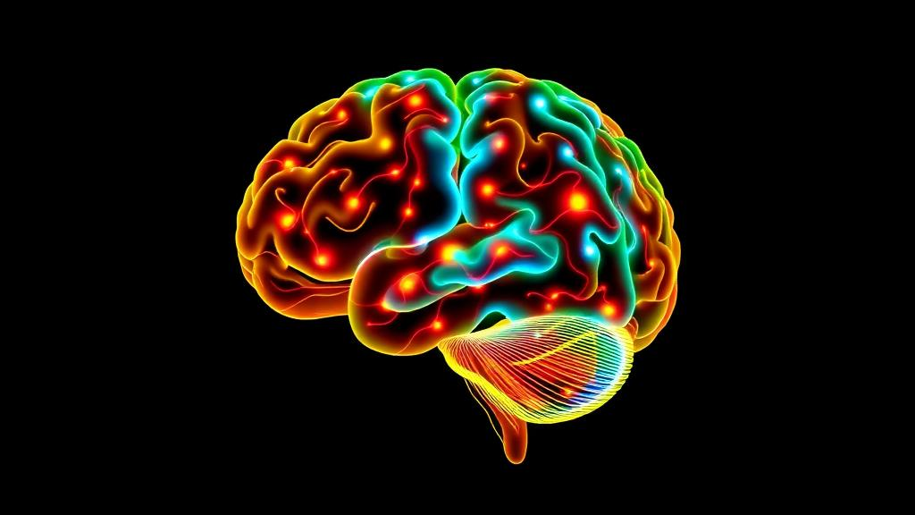
Every time you learn a new movement—whether it is a tennis serve, a piano scale, or, in one recent study, an unfamiliar foot-drawing pattern—your brain quietly reorganizes itself. Capturing that reorganization in living humans, however, is notoriously difficult. Functional magnetic resonance imaging offers exquisite spatial detail but cannot follow the millisecond-by-millisecond electrical conversations of neurons. Electroencephalography, or EEG, offers that temporal precision at the bedside, but for decades it has carried a stubborn limitation: the signals recorded on the scalp are blurred mixtures of activity from many brain regions, and untangling them—the so-called inverse problem—has remained one of neuroscience’s hardest computational challenges.
A team at the University of Tehran, led by Melisa Daryayi and Fariba Bahrami, reports in Scientific Reports an enhanced model-based EEG source imaging method that pushes this untangling considerably further. Their framework, described in a paper published on 12 October 2026, combines a multi-layer perceptron neural network with finite element modeling of the human head to estimate each subject’s tissue conductivities, a step that has traditionally been approximated with generic values. The result is a system that can resolve up to six simultaneously active sources across 116 cortical and subcortical regions of the brain, achieving a reported source localization accuracy of 85.98 percent—a substantial leap over the roughly 67.1 percent accuracy reported for existing comparison methods, even when the signals are contaminated with noise.
To understand why this matters, it helps to grasp the physics of the problem. When neurons fire in synchrony, they generate tiny electrical potentials that propagate through the skull and scalp, where electrodes pick them up. But the skull, brain tissue, and scalp all conduct electricity differently, and in every individual those conductivities vary. If a researcher assumes the wrong conductivity values, the mathematical inversion that maps scalp voltages back to brain sources will place those sources in the wrong locations. The Tehran team’s approach attacks this weakness directly: finite element modeling builds a realistic, subject-specific model of how currents flow through the head, while the neural network learns to estimate the conductivity parameters that best explain the recorded data. The two components work in tandem, refining the forward model that the inverse solution depends on.
The method’s ability to separate deep sources from superficial ones is perhaps its most striking feature. EEG is often criticized as a purely cortical technique, because signals from subcortical structures such as the basal ganglia, thalamus, and cerebellum are heavily attenuated and smeared by the time they reach the scalp. To test whether their framework genuinely distinguishes deep from shallow activity rather than letting one bleed into the other, the researchers performed a dedicated leakage analysis using both single-region and multi-region simulations. The analysis confirmed minimal cross-talk between cortical motor sources and deep subcortical and cerebellar sources, supporting the claim that the method can reliably separate activity across different brain depths—a prerequisite for studying motor learning, which is known to involve both cortical networks and deep structures.
With the method validated in simulation, the team turned to a proof-of-concept application: tracking brain reorganization as five healthy adults learned a novel foot-drawing task across multiple sessions. Motor skill learning is a textbook case of experience-dependent plasticity. Early in practice, learners rely on effortful, cognitively controlled movements, and brain activity tends to be broad and distributed. As skill consolidates, activity typically becomes more refined and efficient, with shifts in the balance between cortical motor areas, subcortical loops, and the cerebellum. But documenting those shifts non-invasively, with the temporal resolution to see how they unfold, has required exactly the kind of tool the researchers built.
The behavioral results followed a familiar learning curve: participants’ performance improved noticeably after the fourth session, marking a transition point in skill acquisition. When the researchers applied their source imaging framework to the EEG recordings from each session, they found a broad reorganization of cortical and subcortical activity across the training period, a pattern consistent with the functional refinement of motor networks as practice progressed. In other words, as the foot movements became smoother and more automatic, the underlying brain activity did not simply stay in place—it migrated, redistributed, and reorganized across the network of regions that support skilled movement.
The authors are careful to frame these findings appropriately. With only five participants, the learning-related results are explicitly described as preliminary—suggestive of the dynamic changes that accompany motor learning rather than definitive evidence of them. This is an honest caveat, and it underscores the study’s real contribution: the method itself. The imaging framework is the primary advance, and the foot-drawing experiment serves as a demonstration that the technique can extract meaningful, depth-resolved source dynamics from real EEG data collected during a genuine learning task, not just from idealized simulations.
Still, the implications are considerable. EEG is inexpensive, portable, silent, and safe, which makes it far more scalable than fMRI for studying learning over many sessions, in many populations, and in naturalistic settings. A non-invasive method that can localize activity in deep brain structures with nearly 86 percent accuracy would open doors to studying motor rehabilitation after stroke, the progression of movement disorders such as Parkinson’s disease, skill acquisition in athletes and musicians, and the development of motor control in children—all with a cap of electrodes rather than a scanner tube. The combination of computational modeling with neuroimaging of motor behavior, as the authors put it, offers a practical path toward that goal.
The technical architecture also reflects a broader trend in neuroscience: the fusion of data-driven machine learning with physics-based modeling. Purely data-driven approaches can struggle to generalize because they learn statistical patterns that may not hold for new individuals. Purely model-based approaches depend on assumptions—like generic conductivity values—that may not hold for any particular individual. By using a neural network to personalize the finite element model, and the finite element model to constrain the neural network’s inverse solutions, the framework hedges against the weaknesses of both paradigms. The reported robustness under varying noise conditions suggests that this hybrid strategy pays off precisely where EEG source imaging has historically been most fragile.
For now, the method’s learning-related findings await confirmation in larger cohorts, and the authors themselves flag that limitation. But the study marks a meaningful step toward a long-sought capability: watching the brain rewire itself in real time, at the level of individual cortical and subcortical regions, using nothing more than electrodes on the scalp and a well-designed computational model. If subsequent studies bear out the framework’s accuracy at scale, the humble EEG cap could become a window not just onto the brain’s electrical chatter, but onto the physical process of learning itself—session by session, region by region, as new skills take shape in the circuitry of the mind.
Subject of Research: An enhanced model-based EEG source imaging method for tracking brain reorganization during motor skill learning
Article Title: An enhanced model-based EEG source imaging method captures dynamic changes during motor skill learning
Article References: Daryayi, M., Hassanzadeh, Z., Mahdavian, A., Bahrami, F., Kalhor, A., & Moshiri, B. (2026). An enhanced model-based EEG source imaging method captures dynamic changes during motor skill learning. Scientific Reports. https://doi.org/10.1038/s41598-026-75574-1
Image Credits: AI Generated
DOI: 10.1038/s41598-026-75574-1
Keywords: EEG source imaging, inverse problem, finite element modeling, neural network, motor skill learning, brain reorganization, subcortical sources, cerebellum, neuroplasticity, Scientific Reports, enhanced, model-based
News Source: Cassandra Pierce. (October 14, 2026). Smarter EEG Brain Imaging Tracks How the Brain Rewires Itself as We Learn New Movements. Scienmag.