Credit: Keith Mickolajczyk / Penn State
A toddler running sometimes loses footing because both feet come off the ground at the same time. Kinesin motors that move materials around in cells have the same problem, which limits how fast they can traverse a microtubule in the cell and carry cargo, according to Penn State researchers who have now seen these kinesin motors move using an unusual microscope and tagging method.
"We can now see biological processes at molecular resolution and at sub-millisecond time scales," said William O. Hancock, professor of biomedical engineering and director of the Intercollege Graduate Program in Bioengineering, Penn State. "To understand how motors work on a nanoscale and millisecond scale we need to see how the motors are walking. We know that neurons require transport for them to grow and survive, and materials need to travel from one end (of the neuron) to the other."
Molecular motors, in this case kinesins, are little machines that use chemical energy to generate mechanical forces sufficient to carry materials through the cell. These molecules have two limbs joined together with attachment devices on the ends that researchers call "heads" but would be better thought of as "feet."
"Diseases like Alzheimer's, ALS and others, have defects in the transport process (in neurons) and it is not understood at the molecular level what defects are and how they affect transport," said Hancock. "We think roadblock proteins bind to the microtubule tracks and impede motor movement. We can measure steps at the roadblock, and hopefully it will help us understand the disease states where transport isn't working."
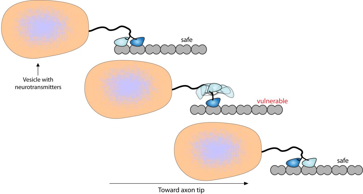
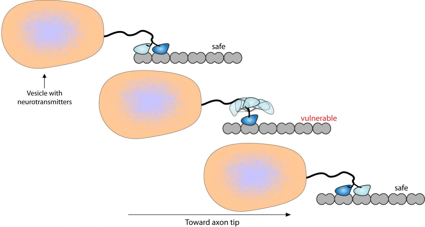
When a kinesin molecule moves from the center of a microtubule toward the end, it is typically carrying cargo. One "foot" attaches to the tubule and then the other back "foot" detaches, swings over and attaches. The molecules are very stable in the short moment when both "feet" are attached, but less so at the time of detachment. The coordination of attachment and release are critical in the molecule successfully walking down the tubule.
Hancock and Keith J. Mickolajczyk, doctoral candidate in bioengineering, note in a recent issue of Biophysical Journal, that "Despite its fundamental importance to the diversity of tasks that kinesins carry out in cells, no existing quantitative model fully explains how structural differences between kinesins alter kinetic rates … to produce functional changes in processivity."
Processivity is the average number of steps the molecular motor can take before it detaches from the microtubule and another must take its place.
Mickolajczyk built the high-resolution, single-molecule microscope so that the researchers could directly see the molecular motor move. To do this, they tagged the molecule on one "foot" with a gold nanoparticle. This allowed the researchers to follow the molecule by reflecting various types of light off the gold.
The researchers found they could model the kinesin movement as a race between attachment of the forward "foot" and detachment of the rear "foot." This walking pattern is governed both by the chemical actions of energy release from adenosine triphosphate — the biological energy storage molecule — and the mechanical push and pull of tethering and untethering. Altering various properties of the kinesin changes the attachment and detachment rates.
Kinesins usually carry their cargo in vesicles — balloon-like, water-filled sacs inside cells. More than one kinesin motor can haul a vesicle at a time, and the motors fall off and are replaced by other kinesin motors during the movement along the tubule. The kinesin "feet" have defined locations on the tubules at which to bind.
"Clarifying these stepping kinetics is very satisfying because these questions have been around for 20 years, but only now do we have the technology to answer them," said Hancock.
###
The National Institutes of Health supported this work.
Media Contact
A'ndrea Elyse Messer
[email protected]
814-865-9481
@penn_state
http://live.psu.edu