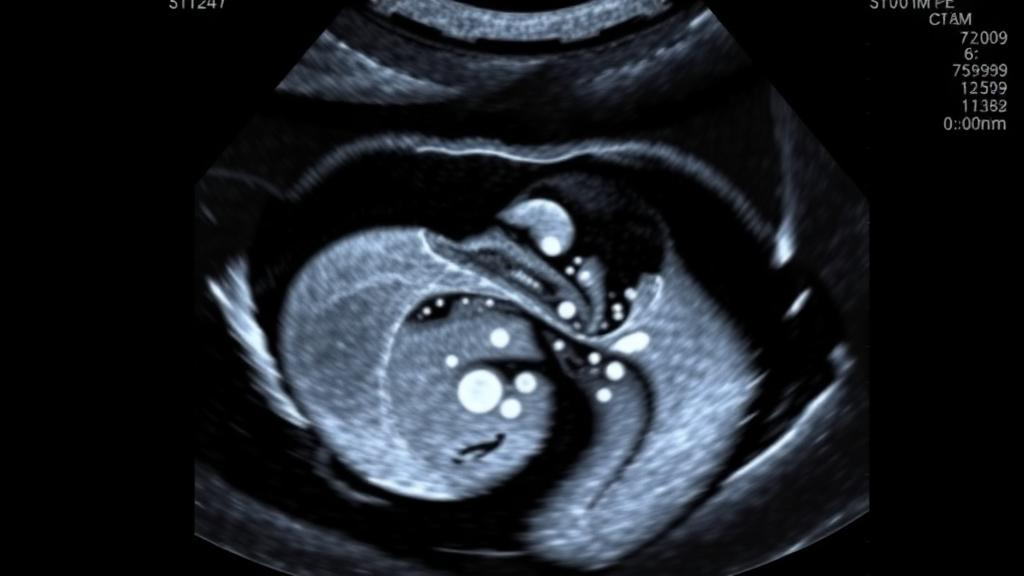
One of the most consequential numbers in prenatal medicine is a simple width measurement. During a routine mid-pregnancy ultrasound, sonographers measure the width of the fluid-filled lateral ventricles in the fetal brain, a value known as the atrial width. When that measurement exceeds roughly ten millimeters, the condition is called ventriculomegaly, one of the most common central nervous system abnormalities detected before birth. The finding matters enormously: enlarged ventricles can signal underlying chromosomal abnormalities, congenital infections, or other structural defects, and the degree of enlargement shapes everything from the recommendation for fetal magnetic resonance imaging to genetic testing and counseling about long-term neurological outcomes. Yet for all its importance, the measurement itself remains stubbornly subjective, dependent on the training, patience, and consistency of whoever holds the ultrasound probe.
A new study published in BMC Medical Imaging by Xiaomei Tang, Huanwen Liang, and colleagues at the People’s Hospital of Guangxi Zhuang Autonomous Region and Shenzhen University argues that artificial intelligence can take that subjectivity out of the equation. The team developed a deep learning framework that automatically measures fetal ventricular width from standard ultrasound images and classifies whether the fetus has ventriculomegaly. In a multi-center evaluation spanning more than a thousand fetuses, their best-performing model reached 98.20 percent diagnostic accuracy and matched expert annotations with a mean absolute error of just over half a millimeter, outperforming radiologists of varying experience levels in consistency, if not always in raw skill.
The technical challenge the researchers confronted is one familiar to anyone who has worked in medical image analysis. Ultrasound is notoriously noisy, with speckle artifacts, acoustic shadows, and ambiguous tissue boundaries. Identifying the atrium of the lateral ventricle and placing calipers correctly requires recognizing anatomical landmarks such as the choroid plexus and the parieto-occipital sulcus, structures that look different depending on fetal position and gestational age. Even experienced sonographers can disagree with one another by a millimeter or more, and because the diagnostic threshold between normal and abnormal sits at ten millimeters, small measurement variability can flip a clinical decision. The researchers designed their study specifically to quantify this problem and test whether a neural network could beat it.
The team assembled ultrasound data from 1,018 fetuses collected across four hospitals, a scale that lends the results unusual credibility for a single-architecture study in this domain. Images from normal fetuses and those with ventriculomegaly were randomly split, with roughly 80 percent of the data used to train the models and 20 percent reserved for internal validation. Crucially, the investigators also curated an independent external validation dataset, collected separately, on which the framework’s performance could be tested against human radiologists ranging from junior to expert. External validation is widely considered the gold standard for evaluating medical AI, because it tests whether a model has genuinely learned the anatomy rather than memorizing the quirks of a particular scanner or hospital population.
Architecturally, the researchers compared two distinct strategies for extracting the ventricular width from an image. The first, called AI-LandVM, framed the task as landmark localization: the network identified specific anatomical points, and the distance between them yielded the measurement. The second, AI-SegVM, took a segmentation-based approach, using a U-Net architecture to trace the full boundary of the ventricle and derive the width from the resulting region. Both strategies rested on the U-Net, a convolutional network with an encoder-decoder structure that has become the workhorse of medical image segmentation. Its encoder compresses the image into abstract feature maps capturing texture and shape, while its decoder re-expands those features back to full image resolution, producing a pixel-level map of what matters in the scene.
The head-to-head comparison produced a clear verdict. The segmentation-based AI-SegVM framework substantially outperformed the landmark-based alternative, and the authors attribute the difference to a fundamental asymmetry in what each strategy can see. A landmark approach reduces an entire anatomical structure to a handful of coordinate points, discarding most of the spatial context of the ventricle. Segmentation, by contrast, forces the network to understand the complete morphology of the structure, its boundary, its extent, and its relationship to surrounding tissue, before any measurement is made. In effect, the richer supervisory signal of a pixel-wise mask teaches the network a more robust internal representation of fetal neuroanatomy, which translates into more reliable measurements when faced with the variability of real-world scans.
On the external validation set, AI-SegVM delivered impressive numbers. Its mean absolute error against expert annotations was 0.53 plus or minus 1.17 millimeters, meaning the average deviation was small and the distribution of errors remained tightly controlled. The intra-class correlation coefficient, a statistical measure of agreement that ranges from zero to one, reached 93.02 percent, indicating near-excellent concordance with expert measurements. Diagnostic accuracy stood at 98.20 percent. Just as important was what the Bland-Altman analysis, a standard technique for assessing agreement between two measurement methods, revealed about consistency. The model’s measurements clustered tightly around expert values without systematic bias, while measurements taken by radiologists with less experience showed wider scatter and greater variability, exactly the kind of operator dependence the study set out to address.
The comparison with human readers deserves particular attention. Rather than simply benchmarking the AI against a single expert consensus, the researchers evaluated how radiologists of different skill levels performed on the same images. The result was a portrait of the variability problem in miniature: more experienced readers produced measurements closer to expert ground truth, while less experienced readers deviated more, particularly on borderline cases where the ventricular width hovered near the diagnostic threshold. The AI system, by contrast, delivered consistent performance regardless of case difficulty, effectively behaving like an expert reader whose accuracy never degrades with fatigue, workload, or limited exposure to rare presentations. For the authors, this consistency, as much as raw accuracy, is the key clinical argument, since standardized measurement is precisely what screening programs need to reduce false positives and false negatives alike.
The study, supported by the Guangxi Key Research and Development Program and the Guangxi Natural Science Foundation, was approved by the ethics committee of the People’s Hospital of Guangxi Zhuang Autonomous Region and conducted in accordance with the Declaration of Helsinki, with informed consent waived due to its retrospective design. The authors report no competing interests, and the article is published open access under a Creative Commons license. As with any retrospective study, the findings represent a carefully controlled evaluation rather than proof of real-world performance, and the framework would need prospective testing before clinical deployment. Nevertheless, the work illustrates a broader trend in medical imaging AI: rather than replacing clinicians, deep learning systems are increasingly positioned as calibration tools, standardizing tasks where human performance varies and giving every patient, regardless of which hospital or which sonographer performs the scan, a measurement governed by the same consistent standard.
Subject of Research: Deep learning for automated measurement and diagnosis of fetal ventriculomegaly in prenatal ultrasound
Article Title: Automated measurement and diagnosis of fetal ventriculomegaly in ultrasound using deep learning
Article References: Tang, X., Liang, H., Zhu, X., Liang, S., Yuan, M., Wei, L., Liu, W., Li, X., Liao, S., Zheng, S., Xu, Y., Ni, D., & Zheng, H. (2026). Automated measurement and diagnosis of fetal ventriculomegaly in ultrasound using deep learning. BMC Medical Imaging. https://doi.org/10.1186/s12880-026-02854-7
Image Credits: AI Generated
DOI: 10.1186/s12880-026-02854-7
Keywords: fetal ventriculomegaly, prenatal ultrasound, deep learning, U-Net, image segmentation, landmark localization, Bland-Altman analysis, medical imaging AI, lateral ventricle measurement, diagnostic accuracy, BMC Medical Imaging, operator variability
News Source: Ophelia Keating. (October 10, 2026). Deep Learning Model Matches Experts at Measuring Fetal Brain Fluid in Ultrasound. Scienmag.