Ovarian cancer remains one of the most lethal gynecological malignancies in the world, and the reason is grimly familiar to oncologists: most patients are diagnosed only after the disease has already spread beyond the ovary. Because early symptoms are vague or absent, the crucial diagnostic moment often arrives when a woman with an ovarian mass is already on the operating table. What surgeons do next depends on whether that mass looks benign or malignant, and getting that call wrong carries serious consequences. A new study published in BMC Medicine suggests that a surprising physical measurement, the way blood serum behaves when it is heated, could give clinicians a far more reliable answer before the first incision is ever made.
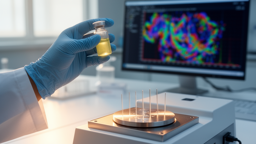
The research, led by Natalia Abian-Franco and F. Javier Falcó-Martí of Hospital Reina Sofía in Tudela and the Institute of Biocomputation and Physics of Complex Systems at the University of Zaragoza, together with senior authors Adrián Velazquez-Campoy and Olga Abian, combined a technique called Thermal Liquid Biopsy with machine learning to classify ovarian tumors as benign or malignant before surgery. The idea behind the approach is deceptively simple. When blood serum is heated in a controlled way using differential scanning calorimetry, the proteins and other molecules suspended in it unfold at characteristic temperatures, producing a thermal fingerprint known as a thermogram. Because cancer changes the composition of the blood, including the abundance and modification states of abundant proteins such as albumin, malignant disease leaves a detectable imprint on that fingerprint.
Differential scanning calorimetry has long been a staple of biophysics laboratories, where it is used to measure the stability of proteins and the binding of drugs to their targets. Applying it to clinical diagnostics is a more recent development, and this study is one of the most thorough attempts to push the technique toward real-world use in oncology. The team analyzed serum samples from 312 patients with a range of ovarian pathologies, drawing on a prospective cohort of 242 patients recruited between 2018 and 2021 and a retrospective cohort of 70 patients whose samples dated from 2011 to 2018. Across the pooled dataset, 186 tumors were benign and 126 were malignant, a distribution that reflects the genuine clinical challenge: in a typical gynecology service, benign masses outnumber malignant ones, which makes it easy for a diagnostic test to look good simply by defaulting to the common answer.
That class imbalance shaped the design of the study in an important way. The researchers built two predictive models. The first, called the OP-only model, was trained exclusively on the prospective samples, giving the most honest and conservative estimate of how the approach would perform on newly recruited patients. The second, an exploratory enriched model designated OP plus OV, added the retrospective samples to the training data, deliberately enriching the malignant class to see whether more cancer examples would sharpen the algorithm’s discrimination. Both models integrated features extracted from the thermograms with clinical and biochemical variables, including established markers such as CA125 and HE4, into what the authors call an integrated TLB and clinical model, or iTLB plus iClin.
The results were striking. The conservative model, trained only on prospective data, achieved an area under the receiver operating characteristic curve of 0.85 for detecting malignancy. In practical terms, an AUC of 0.85 means the model distinguished benign from malignant tumors substantially better than chance and better than many existing single-marker approaches, though it still leaves room for error in individual cases. The enriched model, combining prospective and retrospective data, reached a numerically higher AUC of 0.91 and showed improved classification of indeterminate cases, the gray zone where imaging and biomarkers alone leave clinicians genuinely uncertain. The authors are careful, however, to flag a critical caveat: the retrospective cohort contained malignant cases only, which means the enriched model’s apparent advantage could partly reflect the composition of its training data rather than a genuine leap in diagnostic power.
This kind of methodological honesty matters, because diagnostic studies in oncology have a long history of promising results that fade on contact with clinical reality. The Zaragoza team took several steps to guard against the most common failure modes. They evaluated performance using nested cross-validation, a rigorous scheme in which model tuning and performance estimation are kept strictly separate, reducing the risk of optimistic bias. They also applied regularization techniques, which constrain the complexity of the machine learning models and reduce overfitting, the phenomenon in which an algorithm memorizes the quirks of its training set rather than learning generalizable patterns. Even so, the authors state plainly that external validation in independent, multicenter cohorts is a mandatory prerequisite before any clinical implementation can be considered.
The appeal of Thermal Liquid Biopsy lies partly in what it does not require. Conventional liquid biopsies hunt for tumor DNA, circulating tumor cells, or specific molecular markers, all of which demand that the tumor shed identifiable material into the bloodstream in detectable quantities. Thermal profiling, by contrast, reads a global physical property of the serum, capturing the aggregate consequence of the many molecular changes that cancer induces rather than betting on any single molecule. That makes it conceptually robust to the heterogeneity that makes ovarian cancer so difficult: different tumors, and even different regions of the same tumor, can drive malignancy through different molecular routes, yet all of them perturb the serum proteome in ways that shift the thermal curve. Combined with clinical variables such as age, body mass index, and standard biomarkers, the thermogram gives the machine learning algorithm a rich, multidimensional picture of each patient.
The clinical stakes of better preoperative classification are considerable. When a woman presents with an ovarian mass, the surgical plan diverges sharply depending on the suspected diagnosis. A likely benign mass may be handled by a general gynecologist with a straightforward, fertility-preserving procedure, while a likely malignancy calls for a gynecologic oncologist, often with full surgical staging, and ideally at a specialized cancer center. Misclassification in either direction is costly: benign tumors overtreated with radical surgery expose patients to unnecessary morbidity, while cancers understaged at a first operation can require repeat surgery and may compromise oncological outcomes. Current tools, including ultrasound scoring systems and biomarkers such as CA125 and the risk of malignancy index, leave a meaningful fraction of cases indeterminate, which is precisely where the enriched model in this study showed its most interesting gains.
There are also practical advantages in cost and workflow. Differential scanning calorimetry instruments are standard equipment in biophysics labs, the assay requires only a small volume of serum, and the measurement itself is fast and label-free, requiring no antibodies, amplification chemistry, or sequencing. Turning raw thermograms into diagnostic predictions does require computational infrastructure and validated models, but that is a solvable engineering problem once the clinical validity is established. The Spanish group’s work, funded by the Spanish Ministry of Science, Innovation and Universities, the Instituto de Salud Carlos III, and regional and charitable funders, was conducted with institutional ethics approval and written informed consent from all participants, with samples supplied through the Navarrabiomed Biobank under a formal transfer agreement.
For now, the message from the study is one of cautious optimism. The conservative AUC of 0.85 from purely prospective data establishes that thermal fingerprints carry real diagnostic information about ovarian tumors, and the exploratory results hint that larger, better-balanced training sets could push performance higher. But the path from a promising single-center model to a tool used in operating theaters runs through the unglamorous work of multicenter validation, standardization of sample handling, and prospective trials in genuinely independent populations. If those steps succeed, the idea that a simple heating curve drawn from a tube of blood, interpreted by an algorithm, could help decide how a woman’s cancer surgery is planned may move from biophysics curiosity to standard of care, and ovarian cancer’s devastating pattern of late detection may finally acquire a new line of defense.
Subject of Research: Machine learning analysis of serum thermal profiles for preoperative diagnosis of malignant ovarian tumors
Article Title: Integration of machine learning with thermal liquid biopsy for preoperative assessment of malignant ovarian tumors
Article References: Abian-Franco, N., Falcó-Martí, F. J., Hermoso-Durán, S., Sánchez-Gracia, O., Vega, S., Ortega-Alarcón, D., Velazquez-Campoy, A., & Abian, O. (2026). Integration of machine learning with thermal liquid biopsy for preoperative assessment of malignant ovarian tumors. BMC Medicine. https://doi.org/10.1186/s12916-026-05212-0
Image Credits: AI Generated
DOI: 10.1186/s12916-026-05212-0
Keywords: ovarian cancer, thermal liquid biopsy, machine learning, differential scanning calorimetry, liquid biopsy, serum biomarkers, preoperative diagnosis, CA125, HE4, risk stratification, BMC Medicine, predictive medicine
News Source: Ophelia Keating. (October 9, 2026). Machine Learning Meets Thermal Liquid Biopsy to Spot Ovarian Cancer Before Surgery. Scienmag.