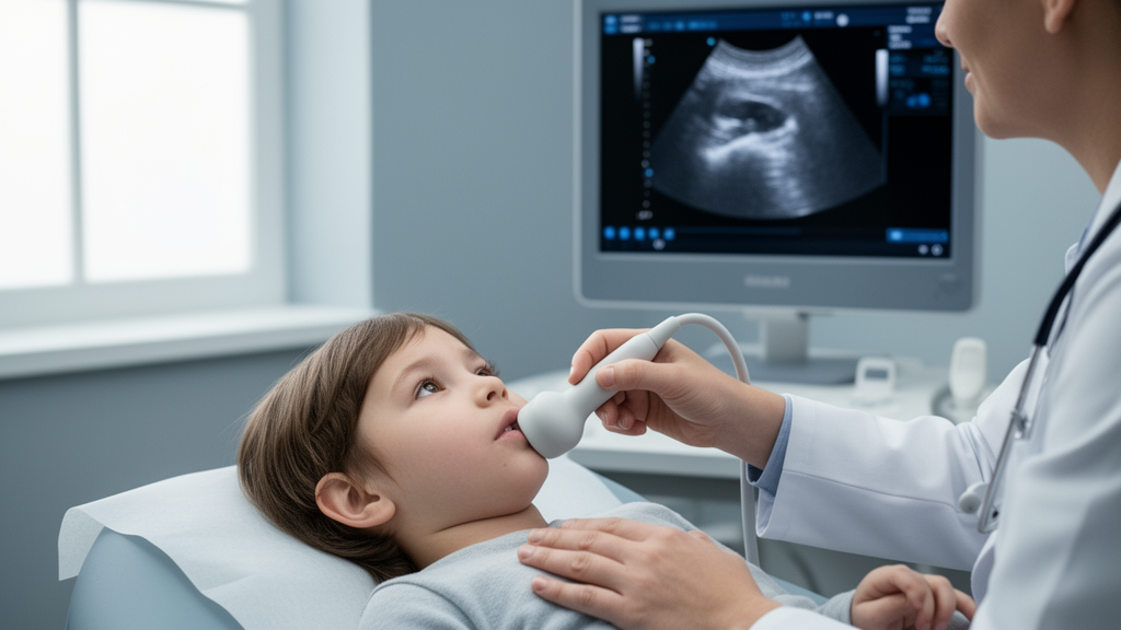
A swollen neck in a child with perfectly normal blood tests has long been one of the more frustrating puzzles in pediatric endocrinology. The gland is visibly enlarged, yet thyroid hormone levels come back normal, and clinicians are left to guess whether the enlargement is a harmless, self-limited phenomenon or the first visible sign of autoimmune thyroiditis, the most common cause of thyroid disease in children and adolescents. A new study from South Korea suggests that the answer may already be sitting on the ultrasound screen. By analyzing nearly a quarter century of pediatric thyroid imaging, researchers found that a simple combination of sonographic abnormalities can identify which children with so-called simple goiter are actually harboring early autoimmune thyroid disease, long before antibodies or hormone shifts make the diagnosis obvious in the laboratory.
The research, published in BMC Endocrine Disorders by a team led by radiologist Sook Min Hwang of Hallym University Kangnam Sacred Heart Hospital in Seoul, examined 88 consecutive children and adolescents who presented with goiter and normal thyroid function between January 2000 and January 2024. Every patient underwent thyroid ultrasonography and biochemical testing for thyroid autoimmunity within three months of each other, allowing the investigators to compare what the ultrasound saw against what the immune system was actually doing. Based on thyroid antibody status, the cohort was divided into two groups: 34 children who tested positive for autoimmune thyroiditis and 54 who did not. The design is elegant in its simplicity, because it isolates a question that clinicians face every day: when a child has a goiter but a euthyroid blood panel, does the ultrasound add anything that blood work cannot yet reveal?
The answer, according to the data, is a resounding yes. Three ultrasound features separated the two groups with striking clarity. Hypoechogenicity, meaning the gland appears darker than normal on the scan, was present in 41.2 percent of the autoimmune group but only 3.7 percent of the non-autoimmune group. Heterogeneous echotexture, a patchy, uneven appearance of the thyroid tissue, was seen in 85.3 percent versus 33.3 percent. And increased vascularity, detectable with Doppler imaging as heightened blood flow through the gland, appeared in 76.5 percent versus 25.9 percent. All three differences were statistically significant at p less than 0.001, a level of consistency across independent imaging features that is rare in studies of this size and speaks to a genuine biological signal rather than statistical noise.
Each of these findings has a plausible pathophysiological explanation rooted in what autoimmune thyroiditis actually does to thyroid tissue. In Hashimoto thyroiditis, the most common form of the disease in children, lymphocytes infiltrate the gland and progressively destroy the follicular cells that produce thyroid hormone. This inflammatory infiltration disrupts the uniform architecture of healthy thyroid tissue, which is why the gland takes on a heterogeneous, pseudo-lobulated appearance. The destruction of follicular cells and their colloid-rich interior reduces the number of acoustic interfaces that normally reflect ultrasound waves, making the tissue appear darker, or hypoechoic. Meanwhile, the inflammatory process triggers angiogenesis, the growth of new blood vessels to supply the immune cell infiltrate, which manifests as increased vascularity on Doppler examination. In Graves disease, the other major autoimmune thyroid condition, stimulating antibodies drive hyperactivity of the gland, which also produces hypervascularity and diffuse textural change.
The most clinically actionable result emerged when the researchers combined these features. When a child showed two or more of the three sonographic abnormalities, the composite criterion achieved a sensitivity of 85.3 percent for autoimmune thyroiditis. In multivariate logistic regression analysis, the composite criterion remained an independent predictor, with an odds ratio of 4.63 and a 95 percent confidence interval of 1.39 to 15.44, corresponding to a p value of 0.013. In practical terms, a child with a goiter and normal thyroid function who displays at least two of these ultrasound findings is more than four and a half times as likely to have autoimmune thyroiditis as a child whose gland looks uniform, normally echogenic, and normally vascular. That kind of risk stratification, derived from an imaging study that is already being performed, costs nothing extra.
The clinical context matters enormously here. Autoimmune thyroiditis is the leading cause of diffuse thyroid disease in the pediatric population, and if it goes undetected it can seriously impair growth, pubertal development, bone mineralization, and cognitive function. The paradox that complicates early diagnosis is that many children present with goiter while their thyroid function remains normal, a state sometimes called euthyroid goiter. Standard practice in many settings is to reassure these patients and monitor them, reserving antibody testing for those who develop abnormal hormone levels. But antibodies can lag behind tissue changes, and the study’s data suggest that the gland often announces the autoimmune process sonographically before the blood test confirms it. Waiting for the laboratory to catch up may mean missing a window in which intervention or closer surveillance could prevent complications.
Beyond the imaging findings themselves, the study identified clinical variables that independently associated with autoimmune thyroiditis. Children in the autoimmune group were significantly older, with a mean age of 14.29 years compared with 11.56 years in the non-autoimmune group, a difference that was statistically significant at p equal to 0.01. They were also significantly more likely to have a family history of autoimmune thyroid disease, at 26.5 percent versus 9.3 percent, with p equal to 0.03. Both findings align with the known epidemiology of the condition: autoimmune thyroiditis has a strong genetic component, with familial clustering well documented, and its incidence rises through childhood and peaks in adolescence, particularly in girls. Age and family history therefore serve as useful contextual clues, but they are far less discriminating on their own than the ultrasound features, which showed much larger absolute differences between the groups.
The authors are careful to frame the composite criterion as a risk stratification tool rather than a definitive diagnostic test, and that framing is scientifically appropriate. Sensitivity of 85.3 percent means that roughly one in seven children with autoimmune thyroiditis would still be missed by the two-abnormality threshold, which is why the researchers position it as a trigger for expedited serological testing rather than a replacement for it. A negative ultrasound pattern, with fewer than two abnormalities, should not end the workup in a child with a concerning presentation. Moreover, the study was retrospective and conducted at a single institution, and the authors explicitly state that prospective multicenter validation is needed before the approach is widely implemented. Ultrasound interpretation also carries an inherent operator dependency, and the prevalence of autoimmune thyroiditis in the study cohort, nearly 39 percent, may be higher than in broader primary care populations, which would affect the predictive values of the criterion in routine practice.
Even with those caveats, the implications for clinical workflow are substantial, particularly in resource-limited settings where every antibody panel represents a real cost. Thyroid ultrasonography is painless, involves no ionizing radiation, and is already the standard first-line imaging modality for pediatric goiter in most guidelines. Embedding a structured assessment of echogenicity, echotexture, and vascularity into the routine interpretation of these scans, and using the two-or-more-abnormality rule to prioritize which children receive immediate antibody testing, could shorten the diagnostic odyssey for many families. It could also reduce unnecessary testing in children whose glands look entirely normal, freeing resources while sparing them the anxiety of prolonged uncertainty. The approach fits into a broader trend in pediatric imaging toward using routinely acquired data to extract earlier, more actionable signals.
What makes the study resonate beyond its immediate clinical utility is the picture it paints of disease unfolding in real time. The thyroid gland of a fourteen-year-old with early Hashimoto thyroiditis is not simply a normal organ that happens to be large; it is an organ under immunological siege, darker and patchier on the scan, flushed with the extra blood flow of active inflammation, all while the endocrine system compensates well enough to keep hormone levels within reference range. The ultrasound, in this light, functions as a window into the earliest phase of the autoimmune process, capturing the tissue-level battle that blood tests will only register once the gland’s functional reserve begins to fail. For the children in this study, that window translated into earlier identification and, potentially, earlier protection of growth and development. For clinicians everywhere who face the daily question of what to do with a child’s painless neck swelling and a normal lab panel, the message is that the image on the screen deserves to be read not just for size, but for texture, brightness, and flow, because those details may hold the diagnosis the blood has not yet revealed.
Subject of Research: Thyroid ultrasonography for early detection of autoimmune thyroiditis in euthyroid children and adolescents with simple goiter
Article Title: Thyroid ultrasonography as an early diagnostic tool for autoimmune thyroiditis in children and adolescents with simple goiter
Article References: Thyroid ultrasonography as an early diagnostic tool for autoimmune thyroiditis in children and adolescents with simple goiter. (n.d.). https://doi.org/10.1186/s12902-026-02432-6
Image Credits: AI Generated
DOI: 10.1186/s12902-026-02432-6
Keywords: autoimmune thyroiditis, thyroid ultrasonography, pediatric goiter, Hashimoto thyroiditis, Graves disease, hypoechogenicity, thyroid vascularity, risk stratification, thyroid antibodies, pediatric endocrinology, early diagnosis, BMC Endocrine Disorders
News Source: Ophelia Keating. (October 9, 2026). Simple Ultrasound Rule Flags Hidden Thyroid Disease in Kids With Goiter. Scienmag.