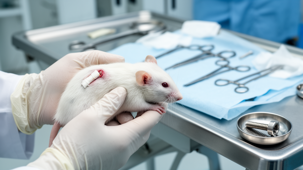
When a titanium implant goes into a bone, it enters a race against bacteria. In the first hours and days after surgery, the implant surface is bare, the surrounding tissue is traumatized, and the local immune defenses are in disarray. Free-floating, or planktonic, bacteria can stick to the metal and begin building a biofilm, a slimy, structured community that shrugs off antibiotics and hides from immune cells. But there is a second, less appreciated scenario: contamination that arrives weeks after the operation, when the wound has healed, fibrous tissue has encapsulated the implant, and the host has partially reasserted control. A new study from researchers at University Medical Center Utrecht, Delft University of Technology, and Utrecht University has now built and validated a single rat model that captures both windows of vulnerability, and in doing so has quantified exactly how much harder it is to seed an infection once the body has settled around an implant.
The work, published in the Journal of Bone and Joint Infection, addresses a stubborn gap in orthopedic research. Implant-associated bone infections are among the most feared complications of joint replacement and fracture fixation, often forcing revision surgery, prolonging morbidity, and driving up costs. Staphylococcus aureus, the culprit in the new study, is particularly troublesome because it adheres readily to biomaterial surfaces and establishes persistent biofilms. Although antibacterial coatings and antibiotic-releasing materials are advancing rapidly, their clinical translation depends on preclinical models that faithfully reproduce the conditions under which infections take hold. Existing animal models vary enormously in implant design, bacterial strain, inoculation route, dose, and timing, making it difficult to compare results across laboratories or to know whether a coating that works in one construct will work in another.
The Utrecht-led team designed a demanding but clinically relevant construct. Adult male Wistar rats, twelve weeks old and skeletally mature, received a six-millimeter segmental defect in the mid-shaft of the femur, created with a saw guide and wire saw. A three-dimensional-printed porous titanium implant was press-fit into the gap, and the whole construct was stabilized with a polyether-ether-ketone, or PEEK, plate fixed by six screws. This is not a simple intramedullary pin or transcortical screw model. The segmental defect creates a large implant-tissue interface, a structured porous scaffold surface, and a complex mechanical environment that mirrors the settings where infection risk is highest clinically: tumor resection, severe trauma, and revision surgery. The porous titanium was chosen deliberately, because its open-pore geometry resembles that of load-bearing porous implants used in real bone reconstruction and provides an ideal substrate for future testing of antibacterial coatings.
Three experiments, involving twenty-eight animals in total, of which twenty-five entered the final analysis, mapped the infection landscape across two time points. In the early model, planktonic S. aureus, a clinical isolate originally obtained from a patient with chronic osteomyelitis, was inoculated directly onto the implant surface at the moment of surgery, at either ten thousand or one million colony-forming units per rat. In the delayed model, the same doses were delivered twenty-eight days after implantation, injected percutaneously into the implant site. A third experiment escalated the delayed challenge to one hundred million colony-forming units, delivered either as free-swimming planktonic bacteria or as a ruptured-biofilm inoculum, in which biofilms grown in the laboratory were scraped from culture wells and dispersed by sonication. Fourteen days after each inoculation, the animals were euthanized and bacterial burdens were quantified in homogenized bone and in sonicated implants, plates, and screws, with micro-computed tomography confirming hardware positioning and implant integrity.
The results were strikingly asymmetric. In the early setting, both doses produced consistent infections across every sampled compartment. Bones carried bacterial loads of roughly one hundred thousand to one million colony-forming units, implants were colonized at around one thousand to ten thousand units, and plates and screws harbored about one hundred thousand units each. Uninfected controls receiving phosphate-buffered saline showed no growth, and the contralateral, unoperated femora remained sterile, confirming that infection stayed localized to the inoculated defect. In the delayed setting, however, the same ten thousand and one million unit doses largely failed. Bone cultures were only sporadically positive, and not a single implant in either delayed group showed detectable colonization, with take rates of zero out of three. The peri-implant environment that had matured over four weeks simply refused entry to bacterial challenges that would have been devastating on the day of surgery.
Escalation changed the picture. When the delayed challenge was raised to one hundred million colony-forming units, delivered by surgically re-exposing the implant, both the planktonic and the ruptured-biofilm groups developed sustained bone infection, with detectable bacteria in every animal. Notably, the ruptured-biofilm inoculum produced higher bacterial burdens on the implant, plate, and screws than the equivalent planktonic dose, while bone burdens were consistently high in both arms. The authors interpret this as evidence that biofilm-derived aggregates carry enhanced infectious potential: the extracellular polymeric substance that shields bacteria from clearance also promotes surface adhesion, and biofilm physiology alters bacterial surface and virulence profiles in ways that favor attachment and immune evasion. Clinically, this matters because although hematogenous seeding often begins with planktonic cells, biofilm fragments shed from distant sites can recolonize implants and sustain chronicity.
Why is a four-week-old implant site so much more resistant? The study’s explanation centers on the maturation of the host-implant interface. At implantation, the surface is unencapsulated, the acute inflammatory response has not yet contained contaminants, and disrupted local defenses strongly favor bacterial adhesion, a dynamic captured by the well-known concept of the race for the surface. By four weeks, wound closure, peri-implant fibrous tissue and callus, and an established local immune milieu create physical and immunological barriers. Only a challenge above a critical threshold, in this case one hundred million colony-forming units, could disturb that equilibrium. Importantly, no animals in the high-dose delayed groups showed signs of systemic illness during the fourteen days after inoculation; body weight losses stayed within two to five percent, and no animal reached the predefined humane endpoints for sepsis, suggesting the infection remained localized rather than spreading.
The authors are candid about the limitations of a feasibility study. Group sizes of three to four animals mean statistical power is low, and the findings should be read as descriptive trends rather than validated reproducibility; formal inferential statistics were deliberately not applied. The one-hundred-million-unit dose required for delayed infection is substantially higher than typical hematogenous bacterial loads, leaving open the question of whether the delayed model best represents late surgical contamination, hematogenous seeding, or a stress test of host defense. Micro-computed tomography was used only qualitatively to confirm hardware position, and infection confirmation relied on microbiological culture without histopathology, so future work should add quantitative bone imaging and tissue-level verification of biofilm architecture. The team also notes that surgical re-exposure in the third experiment, while ensuring accurate inoculum placement, may transiently weaken local barriers compared with percutaneous injection, a potential confounder when comparing across experiments.
Even with those caveats, the model fills a specific and important niche. It allows antibacterial implant coatings to be evaluated under two distinct host-implant maturation states within a single standardized construct, something simpler intramedullary or non-segmental models cannot fully provide. The small pilot cohorts reflect the exploratory, model-establishment nature of the work and align with the principles of replacement, reduction, and refinement that govern ethical animal research. With larger-scale validation, the dual-time-point platform could become a practical proving ground for the next generation of anti-infective implants, tested against both the bacteria that arrive during surgery and the harder-to-defeat challenges that appear weeks later. For the growing population of patients carrying orthopedic hardware, a reliable way to test prevention against both faces of implant infection is a step toward keeping those devices, and the bones around them, safe for good.
Subject of Research: A rat femoral-defect model for studying immediate and delayed implant-associated bone infections
Article Title: Feasibility of a rat femoral-defect model for immediate perioperative and delayed post-implantation implant-associated bone infections
Article References: Keikhosravani, P., Croes, M., Rahmani, N. R., Stabuli, F., Cecotto, L., Vogely, H. C., van der Wal, B. C. H., Koolen, M., Gawlitta, D., Weinans, H., Khodaei, A., & Amin Yavari, S. (2026). Feasibility of a rat femoral-defect model for immediate perioperative and delayed post-implantation implant-associated bone infections. Journal of Bone and Joint Infection, 11(4), 535-546. https://doi.org/10.5194/jbji-11-535-2026
Image Credits: AI Generated
Keywords: implant-associated infection, Staphylococcus aureus, biofilm, rat model, femoral defect, porous titanium, PEEK plate, osteomyelitis, antibacterial coatings, preclinical model, micro-CT, orthopedic surgery
News Source: Ophelia Keating. (October 8, 2026). Rat Model Cracks the Two Faces of Implant Infection, From Surgery to Weeks Later. Scienmag.