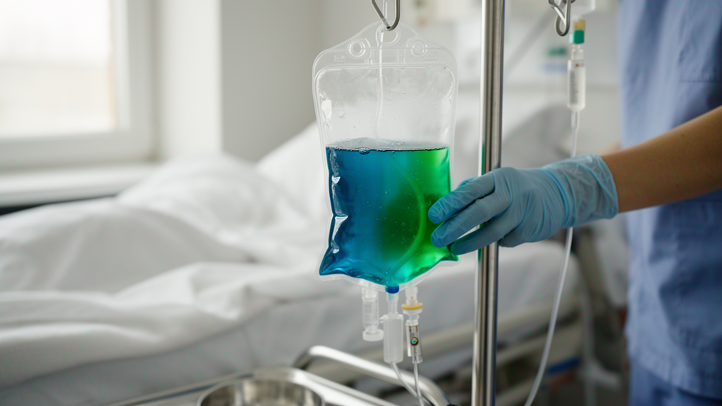
When a 43-year-old woman arrived at the emergency department with burning urination, bacteria in her urine, and an elevated white blood cell count, her care team expected to manage a complicated urinary tract infection. What they did not expect was the sight that greeted them during her admission: the fluid collecting in the tubing of her intraabdominal abscess drain, which had previously been a routine serosanguinous color, had turned a striking blue-green. The case, reported as a clinical image in the Journal of General Internal Medicine by Gabriel Haw of Oregon Health and Science University and Benjamin Vipler of the University of Colorado Hospital, offers a vivid reminder that some of the most dramatic diagnostic clues in medicine are literally colored by the biology of the microbes involved.
The patient’s history was complex. She had suffered abdominal abscesses as a consequence of necrotizing pancreatitis, a severe form of pancreatic inflammation in which devitalized tissue becomes a breeding ground for infection. Those abscesses were being managed with intraabdominal drains, catheters left in place to continuously evacuate infected fluid from the abdominal cavity. On presentation she reported dysuria, painful or difficult urination, and her urinalysis showed evidence of bacteria, while blood tests revealed leukocytosis, an abnormally high white cell count that signals systemic inflammation. Urine culture subsequently grew 100,000 colony forming units of mixed bacteria, a classic quantitative threshold used to support a diagnosis of urinary tract infection.
The blue-green discoloration of the drain output was the detail that transformed an ordinary admission into a diagnostic puzzle. The authors were careful to rule out the most common iatrogenic explanation first: the patient had never received methylene blue or any of the other bodily fluid-bluing medications that can tint urine and drain fluid a similar hue. Methylene blue, used for a range of clinical purposes from treating methemoglobinemia to marking tissues during surgery, is a well-known cause of blue or green discoloration in body fluids, and its exclusion was essential. The team also obtained a CT sinogram, an imaging study in which contrast is injected through the drain tract to map its course, which demonstrated no fistulas, ruling out an abnormal connection between the urinary tract and the drain that might have allowed discolored urine to contaminate the output.
With medication effects and anatomical communication excluded, the blue-green substance from the intraabdominal drain was sent for culture. The result was Pseudomonas aeruginosa, a Gram-negative bacterium famous among microbiologists precisely for the pigments it produces. The color in the drain tubing was not a chemical contaminant or a metabolic oddity of the patient; it was the visible signature of bacterial virulence factors synthesized by organisms colonizing the drain line. In effect, the drain had become a crude culture flask, and the organism had announced itself by changing the color of its surroundings.
The two pigments responsible for Pseudomonas aeruginosa’s characteristic hues have been studied for decades. Pyocyanin, whose name derives from the Greek words for pus and blue, produces the teal to blue-green coloration seen in this patient’s drain output. It is not merely a dye. Pyocyanin is a redox-active phenazine compound that functions as an oxidative weapon in microbial warfare, generating reactive oxygen species that damage competing bacteria and the cells of the host. Its ability to kill or inhibit other microorganisms gives Pseudomonas a competitive advantage in polymicrobial environments, and it contributes directly to tissue injury during genuine infection. The second pigment, pyoverdine, lends a yellow-green fluorescence and serves a completely different purpose: it is a siderophore, a molecule that chelates iron from the environment with high affinity, allowing the bacterium to scavenge this essential nutrient even when iron availability is severely limited, as it is inside the human body.
Blue-green abdominal drain output, the authors note in their discussion, has a defined differential diagnosis. It may be caused by contrast agents, by bacteria, or by discolored urine entering the drain through a bladder fistula. The systematic exclusion of the first and third possibilities left bacteria as the explanation, and culture confirmed which organism was responsible. This diagnostic sequence matters clinically because the implications of each cause differ enormously. A fistula may require surgical intervention, a medication effect requires only drug review, and bacterial colonization of a drain raises the question of whether the organism is truly infecting the patient or simply living on the device.
That distinction, colonization versus infection, became the central management question of the case. In consultation with infectious disease specialists, the team elected to maintain control of the patient’s abscesses with drains alone, without adding antipseudomonal antibiotics. The reasoning was grounded in the patient’s own history and course. Her abscesses had previously not responded to long-term use of antipseudomonal antibiotics, suggesting that escalating antimicrobial therapy again would offer little benefit while risking the well-known harms of broad-spectrum antibiotic exposure, including resistance, toxicity, and disruption of the normal microbiome. More tellingly, her dysuria and leukocytosis resolved without antipseudomonal therapy, a clinical trajectory that strongly suggested the Pseudomonas recovered from the drain represented colonization of the drain line rather than a true pseudomonal abscess infection.
The distinction is subtle but consequential. Pseudomonas aeruginosa is an opportunistic pathogen notorious for its ability to form biofilms on medical devices, including catheters and drains, where encased communities of bacteria are far more tolerant of antibiotics than free-floating planktonic cells. A drain left in an abscess cavity for a prolonged period provides exactly the kind of surface this organism exploits. Yet the presence of an organism in drain output does not automatically mean it is driving disease. In this patient, the systemic signs of inflammation resolved on their own, and source control, the physical evacuation of infected material through drainage, remained effective. The clinical team’s decision to observe rather than treat reflects a growing appreciation in hospital medicine that not every positive culture demands an antibiotic, particularly when the organism may be a passenger on a device rather than an invader of tissue.
For clinicians, the case is a compact lesson in bedside reasoning. An unusual color in a drain should prompt a structured differential: review the medication list for known fluid-bluing agents, consider imaging to exclude fistulous communications, and culture the fluid itself. For the broader public, the story is a striking illustration of how much information can be read from something as simple as the color of a body fluid. The teal hue that alarmed the care team was, in a literal sense, the chemical exhaust of a microscopic competitor, the visible evidence of pyocyanin being deployed in the ongoing war between bacteria for territory and resources. That a physician could look at a drain line and form a reasonable suspicion of Pseudomonas before the laboratory confirmed it speaks to a kind of diagnostic pattern recognition that has existed since the nineteenth century, when early microbiologists first described the blue-green pigment in purulent dressings and named the organism accordingly. Modern medicine has added CT sinograms, quantitative urine cultures, and infectious disease consultation to the toolkit, but the fundamental clue remains the same one visible to any observer at the bedside: the color of the fluid, and the organism that painted it.
Subject of Research: Blue-green discoloration of abdominal drain output caused by pigment-producing Pseudomonas aeruginosa
Article Title: Teal Drain Output
Article References: Haw, G., & Vipler, B. (2026). Teal Drain Output. Journal of General Internal Medicine. https://doi.org/10.1007/s11606-026-10869-7
Image Credits: AI Generated
DOI: 10.1007/s11606-026-10869-7
Keywords: Pseudomonas aeruginosa, pyocyanin, pyoverdine, abdominal drain, necrotizing pancreatitis, urinary tract infection, clinical image, bacterial pigments, drain colonization, infectious disease, source control, antibiotic stewardship
News Source: Ophelia Keating. (October 8, 2026). Blue-Green Drain Fluid Reveals Pigment Warfare of Pseudomonas aeruginosa. Scienmag.